Emergency Department Care
Subungual hematoma
Small (less than 25% of the nail bed [nailbed]) and painless subungual hematomas require no intervention, as the hematoma will eventually reabsorb. If the subungual hematoma covers more than 25% of the nail bed or is causing pain, the patient should be offered evacuation via trephination or nail removal.
Treatment of subungual hematomas covering greater than 25-50% of the nail bed is controversial and varies with personal preference. [19] Historically, treatment includes removal of the nail and repair of any underlying lacerations. This practice came about because 50% of these hematomas have concurrent nail bed lacerations. The incidence of nail bed laceration increases to 94% when associated with a distal phalangeal fracture, regardless of the size of the hematoma. [4, 20, 21, 22, 23]
Studies have shown that as long as the nail is still partially adherent to the nail bed or paronychia and is not displaced out of the nail fold, removal of the nail and repair of the nail bed does not improve outcomes versus simple trephination. Neither the size of the hematoma nor the presence of an associated fracture has been associated with adverse outcomes. [16, 24, 25] Trephination is contraindicated if a fracture requires surgical repair or if the germinal matrix is entrapped within the fracture, as delayed union or the formation of an intraosseous inclusion cyst may occur. [17]
The advantages of simple trephination include less pain for the patient, shorter length of stay, and less costly intervention. [16]
Evacuation of hematomas
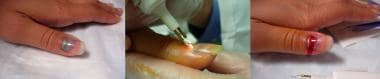
Various methods of trephination exist (shown in the image below). The easiest and safest is to use an electric cautery, which melts a hole through the nail. Once the cautery encounters the underlying hematoma, the tip cools, preventing further injury to the nail bed. If the hole is of adequate size, blood will drain and relieve some pain and the pressure sensation for the patient.
A paper clip may also be used after it is heated until red hot. [26]
An 18-gauge needle may be used by twirling the needle back and forth with slight downward pressure until dark blood return is noted. Use of an 18-gauge needle is less optimal because of the risk of injury to the nail bed once the nail has been penetrated. Alternatively, the needle may be directed at an oblique angle (45-60°) without applying pressure. [27]
Another technique is use of a sterile 29-gauge extra-fine insulin syringe needle. [28] Instead of penetrating the nail, the needle is inserted at the hyponychium parallel to the nail, aimed at the most distal portion of the hematoma. Care is taken to keep the needle closer to the nail versus the nail bed. Once the hematoma is penetrated, the needle may be withdrawn. and light pressure placed on the nail will help with evacuation of the hematoma. This technique may obviate the need for digital block anesthesia and may be favorable in evacuating hematomas of the smaller toe nail beds, where trephination is more difficult.
The use of a 2- or 3-mm biopsy punch has also been described. [29, 30] The biopsy punch is gently twirled back and forth with minimal pressure over the hematoma.
Nail bed repair
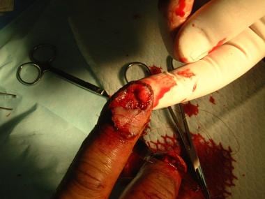
Principles of treatment include minimal debridement, preservation of as much tissue as possible, atraumatic wound care, and splinting with the nail or an alternative material. [31, 32, 13] (Nail bed repair is shown in the image below.)
A digital block of 1% lidocaine hydrochloride without epinephrine provides anesthesia of sufficient duration for most repairs. Bupivacaine extends anesthesia time 4-8 hours for longer procedures.
Children may require procedural sedation and analgesia.
The hand should be prepared with povidone-iodine (Betadine) and covered with sterile drapes. The injured finger should be exsanguinated with a half-inch or 1-inch Penrose drain wrapped in a distal to proximal direction and placed around the base to serve as a tourniquet and provide a blood-free field.
The nail is elevated using the blades of either fine or curved iris scissors or small elevator scissors. Specific care is necessary to not injure the nail bed. A blunt dissecting technique should be used, and the scissors are placed gently underneath the nail until they reach the nail fold. Slowly open the scissors as it is removed. Care must again be taken to avoid further damage to the underlying nail bed or overlying nail fold. Once the nail is sufficiently separated from the nail bed, it is gently removed by applying firm and steady distal traction using a hemostat.
Lacerations to the nail bed should be repaired using 6-0 or smaller absorbable sutures. Minimal to no debridement should be performed because aggressive debridement may cause undue tension on the repair and results in scarring.
When repairing avulsed nails and nail beds, if the nail is detached proximally, it must be removed to inspect for any damage to the nail bed. Careful inspection of the nail is important because often only a fragment of nail bed may be attached to the undersurface of the avulsed nail. Only outer and dorsal surfaces of the nail should be cleaned. Any large fragments of nail bed should be preserved for use as a free graft. Crushing injuries leave many small pieces of nail bed. If all fragments are not incorporated into the repair, they may grow independently and cause nail horns or spicules. If tissue is not available and the defect is small enough, the area will heal effectively by secondary intention.
Simple dorsal roof lacerations can often be repaired by accurately repairing the skin overlying the nail fold. However, if possible, suturing of the dorsal roof with a 7-0 chromic suture may provide more accurate repair. Associated paronychial injuries must be repaired and stented to prevent pterygium or adhesions, as it serves as a mold to coax nail to grow along a proper path. Distal phalangeal fracture reduction and healing is important to final nail formation. Poor reduction of the bone translates directly into irregularities of the nail bed.
The proximal nail should be reinserted into the nail fold. The replaced nail keeps the nail fold open for new nail growth and provides a protective cover for the nail bed and a precise template for new nail to follow as it regenerates. It also serves as a rigid splint for any underlying fractures and reduces postoperative discomfort and improves postoperative function. Some evidence suggests, though, that replacing the nail may be unnecessary [33] and may delay wound healing and increase the risk of infection in children. [34, 35]
Before replacement, a small hole should be made in the nail, preferably so that it is not overlying the laceration. This is to allow drainage and thus prevent a growing hematoma to separate the nail from the nail bed.
The nail is then placed back in the nail fold as a stent and held in position by 5-0 or smaller nylon sutures placed by one or a combination of the techniques below:
-
Distally through the hyponychium and the nail.
-
Through the nail and proximal to the nail fold. [36]
-
Through a half-buried horizontal mattress suture placed down proximal to the nail fold into proximal nail then back out the nail fold.
-
Through the paronychia and nail bilaterally.
-
As a dorsal figure-of-eight suture [37] : A suture is placed transversely just distal to the hyponychium, then placed proximal to the nail fold in the same direction and tied back to itself. If the nail slips laterally, 2 small vertical cuts may be made in the nail for the suture to catch upon.
-
An alternative figure-of-eight suture [38] : A suture is placed along one paronychium for approximately 5 mm, then taken over the nail and sutured along the opposite paronychium in the same direction as the first suture, such that the sutures cross over the nail.
-
Utilizing a double stitch [38] : A combination of both-described figure-of-eight sutures, without necessarily having to create a figure-of-eight suture with each stitch. A suture is placed along one paronychium for approximately 5 mm, then taken over the nail and sutured along the opposite paronychium in the opposite direction as the first suture and tied over the nail. A second suture is then placed proximal to the eponychium and taken over the nail plate through the fingertip just distal to the hyponychium and tied off.
-
In lieu of sutures, tissue adhesives such as Indermil (n- butyl cyanoacrylate) or Dermabond (octyl-2-cyanoacrylate) may be applied along the perionychium after the nail is replaced. [39] Tissue adhesives are also a less invasive option for nail bed and nail repair. [40]
In one randomized controlled trial, nail bed laceration repair using Dermabond required less time with no difference in cosmetic or functional outcomes compared to suture repair. [41] The adhesive should be allowed to dry prior to replacing the nail.
Nail fragments may be repaired together first using adhesive, then secured into the nail fold by a thin layer placed under the nail, using gentle downward pressure while the adhesive dries. [42]
Alternatively, nail fragments may be pieced together on the nail bed, with a light coating of adhesive wiped or dripped between adjoining fragments and on skin adjacent to the perimeter of the nail. As the adhesive dries, use forceps to maintain external pressure. [43]
Chloramphenicol ointment may be used in a similar fashion for simple lacerations, with a small amount applied under the nail so that the ointment forms an adhesive layer as it is positioned into the nail fold. [44]
-
Dress the injured finger with nonadherent gauze and 2-inch gauze roll, then splint the finger.
A hand surgeon should be consulted for significantly avulsed nail matrix or for severe crush injuries.
In general, except for a simple subungual hematoma in which the nail bed was not inspected for potential laceration or injury, a wound check in 2-5 days is suggested to check for infection and to repack the nail fold, if necessary. Sutures should be removed from any replaced nail in approximately 2-3 weeks. If acrylic nail, hypodermic syringe sheath, or other material was used as a stent, it should be removed in 3 weeks. If the original nail was used as a splint, it will be pushed out as new nail grows in, and it will fall out on its own.
In a study by Chiche et al, glue (2-octylcyanoacrylate) and absorbable sutures were compared for nail bed repair in children (74 nail bed lacerations in 68 children). In the study, 36 nail beds were repaired with glue in the ED versus 38 sutured in the operating room. The rate of nail dystrophy was 14% (5% major) regardless of the technique used, but time of nail bed repair was significantly shorter in the ED glue group (10.2 vs. 20.3 min, P< 0.001). The complication rate was higher for patients treated in the ED. [32]
Surgical Care
Small (less than 25% of the nail bed) and painless subungual hematomas require no intervention, as the hematoma will eventually reabsorb. If the subungual hematoma covers more than 25% of the nail bed or is causing pain, the patient should be offered evacuation via trephination or nail removal.
Treatment of subungual hematomas covering greater than 25-50% of the nail bed is controversial and varies with personal preference. [4, 20, 21, 22, 23, 19]
Some studies have concluded that as long as the nail is still partially adherent to the nail bed or paronychia and is not displaced out of the nail fold, removal of the nail and repair of the nail bed do not improve outcomes versus simple trephination. Neither the size of the hematoma nor the presence of an associated fracture has been associated with adverse outcomes. [16, 24, 25] Trephination is contraindicated if a fracture requires surgical repair or if the germinal matrix is entrapped within the fracture, as delayed union or the formation of an intraosseous inclusion cyst may occur. [17]
Lacerations to the nail bed should be repaired using 6-0 or smaller absorbable sutures. Minimal to no debridement should be performed because aggressive debridement may cause undue tension on the repair and results in scarring. Tissue adhesives are also a less invasive option for nail bed and nail repair. [40]
When repairing avulsed nails and nail beds, if the nail is detached proximally, it must be removed to inspect for any damage to the nail bed.
A new prospective surgical approach has been discussed for large-area defects of the nail bed with distal phalanx exposure, which is a cross finger fascial flap combined with thin split-thickness toe nail bed graft. [48]
Cosmetic surgery: medical tattooing
Medical tattooing is used to restore part of a patient’s physical integrity and may assist in psychological recovery from the physical and/or psychological consequences of disease, surgery, or trauma. Successful application to simulate reconstruction of the nail bed after removal through a surgical avulsion procedure has been reported. [49]
-
Trephination of a subungual hematoma.
-
Nailbed repair.