History
When contact dermatitis is suspected, the history must include a detailed list of environmental exposures. Determine whether the patient has had any exposure to materials such as plants, paints, dyes, cleaning solutions, soaps, and protective gear such as eyewear and athletic gloves. Ask whether any new products or plants are present in the home or during recreational activities. Toys are possible sources of allergen exposure, particularly electronic devices, and a careful history of exposures is needed. [23]
Query patients regarding hobbies that might be the source of an irritant or allergen. Determine whether the patient is applying any products or treatments to the involved area. If the lesions or symptoms appear to be primarily in exposed areas, determine how much sun exposure has occurred recently. Ask the patient if symptoms improve over weekends or vacations.
A detailed history may help determine whether the patient has irritant, allergic, or photo contact dermatitis or contact urticaria. The history should include the following questions:
What is the chief symptom? Irritant contact dermatitis, which is the most common form of contact dermatitis, usually causes mild pruritus or a burning sensation. Allergic contact dermatitis and contact urticaria are usually very pruritic. Pruritus is the main symptom in photoallergic reactions, whereas a burning sensation on affected (ie, sun-exposed) areas of the body is the primary symptom of phototoxic reactions.
When did the symptoms start? If a suspected substance is recognized, how long prior to the symptoms did the exposure occur? With irritant contact dermatitis, symptoms may occur within minutes of the exposure. Mild irritants require prolonged or repeated exposure before inflammation is noted, while strong irritants, such as strong acids and alkalis, can produce an immediate reaction similar to a thermal burn.
Allergic (type IV hypersensitivity) reactions usually take 6-24 hours to produce symptoms. Contact urticaria is usually rapid in onset. Because symptoms occur so rapidly following exposure, the etiology for contact urticaria is usually obvious. If the etiology is not apparent, an exposure history should include items commonly associated with this disorder (see Etiology).
Is this the first time this has occurred? When symptoms are episodic, an accurate diary of exposures occurring shortly prior to symptoms may help narrow the list of possible irritants or allergens.
Has the dermatitis been spreading? Allergic contact dermatitis frequently appears to spread over time. In fact, this represents delayed reactions to the allergens.
Several factors may produce the false impression that the dermatitis is spreading or is contagious. Heavily contaminated areas may break out first, followed by areas of lesser exposure. Thick skin may react much later than thin skin or may not react at all. Different sites may have come in contact with the allergen at different times. Gloves and other clothing contaminated with sap from poison ivy may expose the skin days, weeks, or months later.
The patient's age and the location and appearance of the dermatitis frequently lead the history in a particular direction. For example, if the dermatitis is perioral, the history might include exposure to the following:
-
Pacifiers
-
Bubble gum
-
Musical instruments played with a mouthpiece
-
Toothpaste
-
Mouthwashes
-
Lip-licking habits
-
Sports involving mouthpieces (eg, snorkeling, diving)
-
Lipstick
-
Lip balms
-
Products applied to treat the perioral symptoms
-
Sucking limes and lying in the sun
-
Eating foods such as mangos (specifically, exposure to the skin rind of the mango)
Occasionally, simply asking about some of these possible allergens may stimulate the patient or parent to recall an exposure they had forgotten. It is important to consider allergic contact dermatitis in children with dermatitis that is poorly responsive to therapy or in unusual distributions. [24]
Additionally, certain sites are more likely to be associated with identifiable contact allergen in childhood, including hands, feet, and eyelids.
Photo contact dermatitis usually occurs on sun-exposed areas (at some clothing-optional beaches or in tanning booths, sun-exposed areas may include most, if not all, of the skin surface). A detailed exposure history, including a detailed history of types and quantity of light exposure, is required. Determine whether the reaction occurred following exposure through window glass or on a cloudy day. This would suggest photo dermatitis related to ultraviolet A light.
Although the possible causes of contact dermatitis are virtually endless, identification of the probable type can help direct the search for a provoking agent. For a listing of the more common causes of contact dermatitis, see Etiology.
Physical Examination
Many cases of contact dermatitis have a similar appearance regardless of the mechanism or cause of the inflammation. Other than distribution and severity, most cases of acute irritant contact dermatitis appear similar, and the clinical appearance does not suggest the etiologic agent. However, some distributions are highly suggestive of the etiologic agent. For example, pruritic dermatitis of the ear lobes or near the umbilicus almost always is the result of nickel allergy.
Inflammatory responses can be categorized into acute, subacute, and chronic phases.
In acute contact dermatitis, the skin is bright red and edematous. Clear fluid-filled vesicles or bullae may develop in these areas. As lesions break, they weep clear serum. Yellow crusts form as this serum dries. These may suggest that the area is infected. Although secondary infection can occur, it usually develops over several days and is usually more purulent than the yellow crusts. Most healthy patients do not require antibiotic therapy unless significant purulent drainage is noted or the healing of the wound is not progressing as expected.
Subacute contact dermatitis is less edematous and erythematous. Little or no drainage of serum is present. Superficial papules and excoriations are common.
Chronic contact dermatitis is characterized by scaling, fissuring, and lichenification with minimal edema. Mild erythema and excoriations are common. Pediatric patients of color may develop lichenified lesions, lichenoid papules, and/or postinflammatory hyperpigmentation more often than their white counterparts.
Patients with severe allergies may experience anaphylaxis. Anaphylaxis occurs most commonly in patients who are extremely sensitive to latex and is less common in patients sensitive to exposure to other antigens. Consequently, equipment and gloves should be latex-free during clinical examinations and surgical procedures of patients with whose history suggests extreme sensitivity to latex.
The clinical appearance of the dermatitis may suggest the type of contact dermatitis. This may help to narrow the list of possible causes.
Irritant contact dermatitis
Rash is often localized to the site of exposure. The most common site is the hands.
Severity depends on the irritant, concentration, dwell time, site, and condition of the skin. Thick dry skin is the most resistant to the effects of irritants. Maceration makes skin more vulnerable to irritants. Xerosis can predispose to irritant dermatitis.
Allergic contact dermatitis
In allergic contact dermatitis, skin involvement may extend beyond the borders of the region exposed to the allergen. Edema is generally much more pronounced with allergic contact dermatitis than with irritant contact dermatitis, and vesiculation is more common.
Clues to the allergen suggested by the distribution of the dermatitis include the following:
-
Scalp and ears - Shampoo, hair spray, hair dyes, earrings, eyeglasses, ear plugs, headphones, telephones, bathing caps, ear drops (Cerumenex)
-
Eyelids - Nail polish (transferred by rubbing), cosmetics, contact lens solution, sport goggles, fragrances, metals, neomycin, oleamidopropyl dimethylamine, tosylamide formaldehyde resin, benzalkonium chloride, other preservatives
-
Face - Airborne allergens (eg, poison ivy from burning leaves, ragweed), cosmetics, sunscreens, nose clips, perfumes
-
Lips - Lip balms, lipstick, toothpaste, mouthwash, bubble gum (ie, rosin, cinnamates), nickel in musical instrument mouthpiece, rubber in snorkeling mouthpiece, cane reed in a clarinet, food (eg, mango skin)
-
Neck - Necklaces, perfumes, aftershave lotion (men or women from contact with someone wearing aftershave), rubber or leather straps
-
Trunk - Topical medication, sunscreens, poison ivy, clothing, undergarments (eg, spandex bras, elastic waistbands), metal belt buckles, dive suits
-
Axilla - Deodorant (axillary vault), clothing (axillary folds)
-
Hands - Soaps and detergents, foods, poison ivy, solvents and oils, cement, metals, topical medications, gloves, athletic tape, rubber additives, innumerable occupational exposures, “slime” (a novelty toy)
-
Wrists - Same as hands; watch, watchband, bracelets
-
Genitals - Poison ivy (transferred by hand), rubber condoms, nickel (allergy from a bed-wetting alarm was confused with herpes genitalis and child abuse), feminine hygiene products. One study noted that toilet seat dermatitis is more common than previously recognized and should be considered in children who present with dermatitis that involves the buttocks and posterior thighs. [25] Diaper wipes, diapers, and toilet paper may also be sources of buttock allergic contactants.
-
Anal region - Hemorrhoid preparations (eg, benzocaine, Nupercaine)
-
Lower legs - Topical medication (eg, benzocaine, lanolin, neomycin, paraben), dye in socks, latex/rubber in socks
-
Feet - Rubber, leather, glues, dyes, or nickel snaps in shoes and sandals; topical medications; swim fins; athletic tape
Photo contact dermatitis
Phototoxic photo contact dermatitis is essentially a severe sunburn or an allergic reaction to the sun, with a primary symptom of burning. In photoallergic photo contact dermatitis, manifestations on sun-exposed areas of the body range from sunburns to eczematous dermatitis or hyperpigmentation. Occasionally, aerosolized contactants may produce a similar clinical appearance.
Contact urticaria
Contact urticaria appears as hives or wheals or edematous pale or pink plaques. (See the image below.)
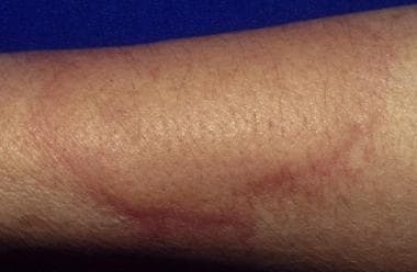 Urticaria, also known as hives or whelps, involves edematous pale or pink plaques. Agents can produce urticaria by immunologic reactions, by nonimmunologic reactions, or by unknown mechanisms. Nonimmunologic reactions are most common. Other types of environmentally associated urticaria must be excluded. This is an example of cold urticaria produced by application of an ice cube to the dorsum of the arm.
Contact reactions to pharmacologically active agent
Typical triple response (ie, erythema, flare, and wheal formation) is noted in seconds. Pruritus may last several hours.
Complications
Risks of contact dermatitis are manifold and include the following:
-
Pain
-
Stigma
-
Stress/psychological distress
-
Scarring/discoloration
-
Superinfection
-
Aggravation of underlying dermatitis (eg, atopic dermatitis)
Secondary bacterial infections are uncommon in the acute stages of contact dermatitis. They may occur several days after damage to the skin and should be treated with systemic antibiotics.
Generalized eruptions secondary to autosensitization may occur in association with severe localized allergic contact dermatitis.
Pulmonary symptoms may occur following inhalation of irritants or potent allergens.
Scar formation may result from deep chemical burns or significant secondary infection.
Phytophotodermatitis usually resolves with extensive postinflammatory pigmentary alteration. Pain and superinfection of blistered regions can also be seen.
In contact urticaria, symptoms of anaphylaxis and systemic reactions can occur with increasing exposures, and sometimes without warning. Therefore, children should strictly avoid known contact allergens and carry an epinephrine autoinjector in the event of severe symptoms with accidental exposure. Patch testing is to be avoided in such cases, owing to the risk of systemic symptoms.
Allergic contact dermatitis in children has significant morbidities, including pain, discomfort, truancy/missed school, itch, disfigurement, and sometimes scarring. For these and many other reasons, early identification and elimination of allergen exposures is important.
Irritant contact dermatitis is often triggered by specific irritants under specific settings, such as the interaction of urine and stool to create irritating products. Recurrence of irritation is not uncommon. In addition, irritation from fragrance contributes to the severity of conditions such as atopic dermatitis. Irritant contact dermatitis can be quite uncomfortable and difficult to manage. Superinfection of irritated skin is not uncommon.
-
Dry, fissured, pruritic eczema is frequently the result of excessive washing and very low humidity in cold climates. Irritant contact dermatitis is due to direct injury of the skin. In this patient, frequent handwashing and use of soap is the cause of damage to the protective layers of the upper epidermis. Patients should be educated about the cause of the dermatitis and instructed in methods of skin protection and care with emollients.
-
Nickel is the most frequent contact allergen in females older than 8 years, and allergy occurs in as many as 25% of females 14 years or older. Allergens, such as nickel, are impossible to completely avoid. Exposure can be reduced with careful instruction, but occult exposures may produce chronic or recurrent symptoms. Nickel in the watch and watch band produced this episode of allergic contact dermatitis.
-
Allergic reactions to rubber products are usually caused by antioxidants and accelerators added in the manufacturing process, rather than the rubber itself. Antioxidants help preserve the rubber, and accelerators help in the vulcanization process. Exposure to rubber in gloves, shoes, undergarments, tires, heavy-duty rubber goods, and sport goggles is common.
-
The typical eruption from poison ivy includes erythema, edema, papules, vesicles, and bullae. Linear streaks as in this patient are characteristic but are not always present. Initial yellow crusts are dried serum from ruptured bullous lesions and not evidence of infection. Oleoresin (urushiol), which exudes from damaged areas of poison ivy, poison oak, and poison sumac, turns black after exposure to air. Fresh oleoresin on the skin dries and may be observed as black smudges or spots.
-
When limes are squeezed into beverages, excess juice remains on the skin. Many other foods can cause similar reactions, i.e. the phytophotodermatitis. Sun exposure of this lime juice produces areas of dermatitis or hyperpigmentation. Perfumes are also common sources of photo contact dermatitis.
-
Most common moisturizers contain various additives and preservatives. The list of ingredients on this bottle is not uncommon, and most of these agents are capable of causing allergic contact dermatitis. Patch testing with dilute concentrations of the individual ingredients can be used to identify the agent that is a problem for any particular patient.
-
Areas of acute contact dermatitis respond well to cool compresses and wound care. Moist compresses are soothing, have a mild antipruritic effect, reduce serous drainage, and gently debride the wound. Clean water, isotonic sodium chloride solution, and Burow solution all can be used. Compresses should be kept moist at all times. Wet-to-dry compresses are painful and destroy fragile tissues. Following moist compress applications for 5-10 minutes, affected sites should be gently cleared of loose crusts and a thin coat of Vaseline or antibacterial ointment should be applied.
-
Urticaria, also known as hives or whelps, involves edematous pale or pink plaques. Agents can produce urticaria by immunologic reactions, by nonimmunologic reactions, or by unknown mechanisms. Nonimmunologic reactions are most common. Other types of environmentally associated urticaria must be excluded. This is an example of cold urticaria produced by application of an ice cube to the dorsum of the arm.
-
Prolonged use of moderate- to high-potency topical steroids may cause skin atrophy or steroid acne. This patient used a moderate-strength steroid, triamcinolone 0.1%, in this area for several weeks. Steroid acne, also called steroid rosacea, has a classic appearance with monomorphic erythematous papules. If the steroid is discontinued, the condition usually worsens. Patients must understand that symptoms worsen before they improve, and several weeks or months are required to taper off this steroid.
-
This purpuric reaction was noted after application of eutectic mixture of local anesthetics (EMLA) for 1 hour. EMLA cream is widely used as a local anesthetic for superficial procedures. Blanching and redness are commonly observed side effects. Dramatic purpuric reactions to EMLA, as in this patient, have been reported. Patch test results in these patients with the individual ingredients of EMLA cream, EMLA cream itself, placebo cream, and Tegaderm are negative. Apparently, the purpuric reaction is not of an allergic nature, but the cream may have a toxic effect on the capillary endothelium.