Imaging Studies
Chest radiographs
Anteroposterior (AP) and lateral chest films are used routinely to assist in the diagnosis of rib fractures, yet sensitivity as low as 50% has been reported. Delayed or follow-up radiographs can be very helpful.
Chest radiographs are much more useful in the diagnosis of underlying injuries, including hemothorax, pneumothorax, lung contusion, atelectasis, pneumonia, and vascular injuries.
Findings of sternal fracture [21] or scapular fracture [24] should increase suspicion for rib fractures.
Rib radiographs
Obtaining a rib radiograph series remains controversial, as the additional information rarely changes the clinical picture or alters treatment. Rib detail radiographs can be helpful in evaluation of the 1st and 2nd ribs and the 7th through 12th ribs. (See the images below.) Formal plain radiographs can also be useful to document abuse for legal purposes.
 Anteroposterior (AP) radiograph of an elderly female patient with severe left chest wall pain after a minor fall. This image demonstrates a left lateral rib fracture (arrow) that is not seen on the standard AP chest radiograph.
 Right rib radiograph in a 48-year-old male who presented with severe right posterior chest wall pain following a fall. This image demonstrates 2 fractures of the right chest wall (white arrows).
Diagnostic sensitivity is higher in rib radiographs than in chest radiographs; however, with a high clinical suspicion, treat for fracture regardless of the radiographic result. In a study of rib fractures in stable adult outpatients presenting with chest pain, a retrospective analysis of chest radiography and a dedicated rib series of 339 patients found that 53 (15.6%) had at least one rib fracture. Only 37.7% of the fractures could be identified on the frontal chest radiograph. The frontal chest radiograph had a sensitivity of 38% and a specificity of 100%. In this series, radiographic findings associated with rib fractures include pleural effusion. Although none of the patients required hospitalization, patients with rib fractures received narcotic analgesia in 47.2% of the cases, significantly more than those without rib fractures (21.3%). [45]
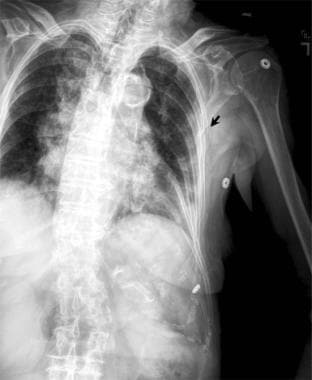
Aortic injury, as shown in the image below, is closely associated with a widening of more than 8 cm measured at the widest points of the mediastinum on an upright anteroposterior chest radiograph.
 Aortic injury is closely associated with a widening of greater than 8 cm measured at the widest points of the mediastinum on an upright anteroposterior chest radiograph.
Clinicians should obtain a skeletal survey in infants suspected of being abused. Findings consistent with abuse include the presence of multiple rib fractures in various stages of healing.
In a retrospective study of 57 pediatric patients, by Pomeranz et al, regarding sensitivity of skeletal survey (SS) radiographs versus CT for diagnosis of rib fractures, 225 rib fractures were identified in 25 patients on CT. and 38 of those fractures were missed on the SS (a miss rate of 17%). [12]
Bedside ultrasonography
Bedside ultrasonography by emergency physicians has been described and provides rapid diagnosis with no radiation exposure. [46]
Advantages include diagnosis of fracture in unossified bone in children and diagnosis of other rib fractures that may be missed on plain radiographs. Small preliminary studies suggest that ultrasonography may be more sensitive than chest radiography in detecting rib fractures when physicians who perform the examination are comfortable using the technique. [47, 48, 49, 50]
Ultrasound also detects costal cartilage fractures and costochondral junction fractures better than radiography. Healing fractures with callous formation and sternal fractures may also be sonographically detected.
The technique includes first clinically identifying the site of maximal tenderness, then using a high-frequency 7-MHz to 12-MHz linear transducer. The transducer is placed perpendicular to the long axis of the rib to identify the posterior surface of the rib by its distinct acoustic shadowing and then rotated 90 degrees to detect any discontinuity of cortical alignment, seen as a break in the hyperechoic rib margin. [46]
Other reported but less common sonographic signs suggestive of fracture include the following: a linear acoustic edge shadow posterior to the fracture, a reverberation artifact posterior to the fracture, and the presence of a hypoechoic hematoma. Once a rib fracture is diagnosed, ultrasonography can then be conveniently and reliably used to exclude the likely complications of pneumothorax and hemothorax. [46]
A limitation is that retroscapular ribs and the infraclavicular portion of the first rib are technically inaccessible by sonography. However, these are uncommon sites for rib fractures. [46]
Chest CT scan
Chest CT scan (see the image below) is more sensitive than plain radiographs for detecting rib fractures. [11] The modality can also provide information regarding the number of ribs involved. [12, 14, 15]
 Axial computed tomography image of the chest in a patient with both anterior and posterior thoracic injuries. A fracture of the sternum (white arrow) and a posterior left rib fracture (yellow arrow) are present.
If complications from rib fractures are suspected clinically or diagnosed by plain radiographs, a chest CT scan may be helpful to document specific injuries, to characterize extent of injury, and to plan for definitive management.
An associated CT scan of the abdomen with intravenous contrast should be considered in cases involving lower rib fractures with suspected or known injury to the liver and/or the spleen. The reported incidence of concomitant intra-abdominal injuries with lower rib fractures is 20-40%. Abdominal CT has been reported to be particularly important if patients are younger than 55 years, have bilateral rib fractures, and have decreased hematocrit levels. [51]
A reduced-dose chest CT scan may detect rib fractures in infants with high suspicion for nonaccidental trauma. In a study of rib fractures in clinically diagnosed cases of child abuse, a retrospective analysis of initial and follow-up skeletal surveys and CT scans of 16 patients younger than 12 months found that 17% (18 of 105) of rib fractures were not documented on the initial skeletal survey. Those fractures not originally detected were seen only after follow-up imaging, more than half of which (11/18) were detected on a subsequent CT. Many of these were posterior (43%) or anterior (30%), and 96% were nondisplaced. [52]
Angiography
Because first and second rib fractures are often associated with vascular injury, ED physicians should consider angiography for such patients, especially if symptoms and signs of neurovascular compromise are present. [7] This is particularly important with posteriorly displaced fractures of the first 2 ribs, which have a much higher degree of association with abnormal angiographic findings than other rib fractures.
While first rib fractures previously were considered a strong risk factor for aortic injury, most authorities now believe that aortography and/or CT scan are not indicated without other evidence of injury, such as abnormal mediastinum.
Bone Scan
A bone scan of the chest wall is the preferred instrument to diagnose rib stress fractures early in the pathologic process. Such fractures typically are not visible on plain films of the chest until very late in their healing, when they develop a visible callus.
Normal costochondral uptake in a child may be intense enough to suggest rib stippling when viewed from a posterior projection.
Any disease or lesion of a rib that results in increased bone turnover may result in positive findings in the ribs.
False-negative results may occur in patients who have recently received iron dextran injections as high levels of iron in the bone marrow interfere with the normal uptake of bone.
MRI
Although MRI is not used as a primary means of detecting rib fractures, displaced or angulated lateral rib fractures as well as posterior rib fractures can be detected by MRI.
Breathing motion can cause artifacts, resulting in nondiagnostic MRIs of anterior rib fractures.
Partial-volume effects may result in a false suggestion of a nondisplaced rib fracture.
Lab studies
Laboratory studies are generally not useful for evaluation of isolated rib fractures. Consider obtaining urinalysis in cases of lower rib fractures, as hematuria may indicate associated renal injury. [43] Tests of the pulmonary function (arterial blood gas measurements) are used to determine if the lungs have been contused but do not actually test for rib fractures.
-
Aortic injury is closely associated with a widening of greater than 8 cm measured at the widest points of the mediastinum on an upright anteroposterior chest radiograph.
-
Anteroposterior (AP) radiograph of an elderly female patient with severe left chest wall pain after a minor fall. This image demonstrates a left lateral rib fracture (arrow) that is not seen on the standard AP chest radiograph.
-
Axial computed tomography image of the chest in a patient with both anterior and posterior thoracic injuries. A fracture of the sternum (white arrow) and a posterior left rib fracture (yellow arrow) are present.
-
Right rib radiograph in a 48-year-old male who presented with severe right posterior chest wall pain following a fall. This image demonstrates 2 fractures of the right chest wall (white arrows).