Approach Considerations
Primary treatment of otitis externa (OE) involves management of pain, removal of debris from the external auditory canal (EAC), administration of topical medications to control edema and infection, and avoidance of contributing factors.
Most cases can be treated with over-the-counter analgesics and topical eardrops. Commonly used eardrops include acetic acid drops, which change the pH of the ear canal; antibacterial drops, which control bacterial growth; and antifungal preparations. Eczematoid (psoriatic) OE often responds to topical steroid drops but may be chronic or recurrent. The ear may require frequent suction debridement under a microscope. If significant canal edema develops, an ear wick may be used to facilitate delivery of topical medications into the medial canal.
In severe cases, oral or intravenous (IV) antibiotic therapy and narcotic analgesics may be required. [18] In the case of necrotizing (malignant) OE, the patient must be admitted to a hospital for IV antibiotic therapy at the discretion of the consulting otorhinolaryngologist. The treatment that is rendered depends on the likely organism, which is best determined by means of Gram staining of the affected area.
The American Academy of Otolaryngology–Head and Neck Surgery Foundation (AAO-HNSF) has published clinical guidelines on the diagnosis and management of OE (see Guidelines). [1]
Removal of Debris From Ear Canal
Removal of debris from the ear canal improves the effectiveness of the topical medication. Gentle cleaning with a soft plastic curette or a small Frazier suction tip under direct vision is appropriate. Irrigation with a mix of peroxide and warm water may be useful for removing debris from the canal, but only if the tympanic membrane is intact. Any water instilled must be removed to keep from exacerbating the condition.
Not uncommonly, children insert a foreign body in their ear canal and do not mention it to their parents. If any pain accompanies purulent drainage, the possibility of a foreign body in the ear canal should be considered. A patient with a foreign body in place will not improve until it is removed. As Noted (see DDx), batteries that have been placed in the ear canal represent a medical emergency and necessitate expeditious diagnosis and removal.
Pharmacologic Therapy
Before antibiotic treatment was recommended for OE, astringents and acetic acid solutions were commonly used to treat this condition. These solutions can be painful to inflamed ear canals and are not generally used today. An aminoglycoside combined with a second antibiotic and a topical steroid (eg, neomycin−polymyxin B−hydrocortisone) used to be the most commonly prescribed topical preparation; however, the neomycin component led to hypersensitivity reactions and ototoxicity in some patients.
Most physicians prescribe topical antibiotic-containing preparations. Otic antibiotic and steroid combinations have shown to be highly successful in treatment, with cure rates of 87-97%. [19] Other agents used include analgesics for pain relief, acidifying solutions, and, in some instances, antipruritics or antihistamines.
Topical medications
Most cases of acute OE respond well to topical treatment. Antibiotic eardrops, with or without a corticosteroid (given to decrease inflammation), are the mainstay of therapy. Topical acidifying and drying agents (given to alter the pH and to inhibit the growth of microorganisms) may be used in mild or resolving cases and are useful in fungal infections. An antifungal agent may be included as necessary.
Mild OE usually responds to the use of an acidifying agent and a corticosteroid. As an alternative, a 2:1 ratio mixture of 70% isopropyl alcohol and acetic acid may be used. For moderate OE, consideration should be given to adding antibacterial and antifungal agents to the acidifying agent and the corticosteroid.
In a systematic review of therapy for OE, Rosenfeld et al demonstrated little overall difference among the various topical agents used to treat this condition [20, 21] ; however, they found that use of a topical steroid alone increased cure rates by 20% as compared with a steroid-antibiotic combination. The combination of oral antibiotics with otic antibiotic solutions has not been shown to improve treatment success rates. [22]
Use of aminoglycoside antibiotic eardrops in the presence of a perforation or ventilation tube may cause problems. Although this is controversial, many otolaryngologists believe that aminoglycoside eardrops may be ototoxic if they enter the middle ear. In these situations, using an alternative, nonototoxic topical preparation (eg, a fluoroquinolone, with or without a steroid) may be safer.
Fluoroquinolones are not associated with ototoxicity, and ofloxacin is safe in cases of a perforated tympanic membrane. One literature review concluded that OE can be safely treated with an otic suspension containing 0.3% ciprofloxacin and 0.1% dexamethasone and that the inclusion of dexamethasone improves treatment success rates. [19] A German meta-analysis found a trend suggesting the superiority of quinolone monotherapy to classic combination regimens comprising a nonquinolone antibiotic plus a steroid. [23] Finafloxacin otic suspension has been approved by the US Food and Drug Administration (FDA) for treatment of acute OE caused by P aeruginosa or S aureus.
Mild fungal infections can usually be treated with an acetic acid solution, whereas more severe cases may have to be treated with a topical antifungal agent, such as 1% clotrimazole.
In the setting of chronic, noninfectious, therapy-resistant OE, a prospective study by Caffier et al demonstrated that daily use of 0.1% tacrolimus cream (administered via an ear wick [see below] that was changed every second to third day) resulted in high rates of resolution (46% through 1-2 years of follow-up) after 9-12 days of therapy. [24] The study also demonstrated longer periods of symptom-free intervals for those who experienced a recurrence.
A systematic review and meta-analysis by Di Traglia et al evaluated the effectiveness of topical nonantibiotic (ie, antiseptic or steroid) treatment against that of topical antibiotic treatment in the management of acute OE. [25] All of the therapies were effective, with no significant differences among them. The authors concluded that the evidence was insufficient to suggest that topical antiseptic or steroid agents are superior or inferior to topical antibiotics.
Ear wick
If the ear canal is severely swollen, an ear wick may be inserted to facilitate the delivery of topical medications (see the image below). The wick may be commercially prepared from a hard sponge material that expands when wet (eg, the Merocel ear wick or the Pope Oto-Wick), cut from a bigger sponge by the physician, or made from narrow gauze (0.25-in. packing works well).
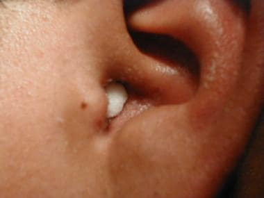 Otitis externa with ear wick in place. Note discharge from canal and swelling of canal.
After the placement of the wick in the ear canal (a process that, unfortunately, causes brief but significant discomfort), the topical antibiotic drops are placed on the external end of the wick to be carried into the recesses of the ear canal. This is done two to four times daily, depending on the recommended dosing frequency for the medication. The wick may fall out as the edema decreases. In any case, it should be removed after 2-3 days.
Analgesics
Pain control is essential to quality patient care. OE can be quite painful, and patients frequently request analgesics. These agents ensure patient comfort and may have sedating properties. Inexpensive, simple nonsteroidal anti-inflammatory drugs (NSAIDs) reduce inflammation and irritation and can be paired with opiates to improve pain symptoms Over-the-counter acetaminophen is appropriate for most patients. In some cases, systemic analgesics are helpful before ear cleaning or wick placement.
Oral and intravenous antibiotics
Most persons with OE do not require oral medications. Oral antibiotics are generally reserved for patients with fever, immunosuppression, diabetes, adenopathy, or an infection extending outside the ear canal. They should be given to individuals with cellulitis of the face or neck skin or to persons in whom severe edema of the ear canal limits penetration of topical agents.
IV antibiotics are used in individuals with necrotizing (malignant) OE; they may also be appropriate for patients with severe cellulitis or persons whose symptoms do not respond to topical and oral antibiotics. A prolonged course of IV antibiotics, lasting for up to 6 weeks, may be required. If the patient is stable, IV antibiotics may be administered at home. Begin treatment with antibiotics to cover pseudomonads, and alter the regimen as necessary on the basis of culture results.
Surgical Debridement and Drainage
Surgical debridement of the ear canal is usually reserved for necrotizing OE or for complications of OE (eg, external canal stenosis). It is often necessary in more severe cases of OE or in cases where a significant amount of discharge is present in the ear. An otolaryngologist usually performs debridement using magnification and suction equipment. Debridement is the mainstay of treatment for fungal infections.
Occasionally, an abscess forms in the ear canal; this usually occurs in cases of OE caused by S aureus. Treatment of the abscess is often accomplished by means of a simple incision and drainage procedure that is usually performed by an otolaryngologist using a needle or a small blade.
Activity
During treatment of OE and for 1-2 weeks after its resolution, advise the patient to keep the ear canal dry. During bathing or showering, advise the patient to place an earplug or cotton ball lightly coated with petroleum jelly in the ear canal to prevent water penetration.
Patients involved in aquatic activities may resume these activities once the infection has been eradicated, generally within 4-5 days. Aquatic athletes may return to the pool earlier than 4-5 days; generally, after 2-3 days of refraining from any water activity, they can return to their usual activities. However, the head must be kept dry until the infection has been eradicated.
The best way of keeping the ear dry, obviously, is to avoid aquatic activities altogether, but the more common practice is simply to limit such activities to those that do not expose the ear to the water (eg, kicking while using a foam floatation board to keep the head above water).
Prevention
Some patients experience multiple recurrences of OE and thus benefit from the adoption of a preventive strategy. The following recommendations related to ear hygiene may help prevent recurrent OE:
-
Eliminate any self-inflicted trauma to the ear canal, such as may occur with the use of cotton swabs or the insertion of objects (eg, bobby pins) into the EAC
-
Avoid frequent washing of the ears with soap; this leaves an alkaline residue that neutralizes the acidic pH of the EAC
-
Avoid swimming in polluted waters
-
Ensure that the ear canals are emptied of water after swimming or bathing; the use of a blow dryer on a low setting after swimming to dry the ear canal has been suggested as a preventive measure, though no studies have demonstrated this suggestion to be effective
-
Instill prophylactic eardrops after each exposure to water to assist in drying and acidifying the ear canal; a combination of 70% isopropyl alcohol and acetic acid in a 2:1 ratio may be used
Some have recommended wearing earplugs for swimming and bathing. If worn, earplugs should be wiped with rubbing alcohol after use. Others have argued that the use of earplugs should be avoided, on the grounds that they may cause trauma to the ear canal and thereby predispose to the development of OE.
Consultations
For simple OE, consultation with an otorhinolaryngologist generally is not necessary. However, such consultation is appropriate if the patient has severe OE, is not responding to treatment as expected, has a suppurative complication or a perforated tympanic membrane, or is suspected of having necrotizing (malignant) OE. Debridement of the ear canal is often necessary for resolution of the infection.
Necrotizing OE necessitates consultation with an otorhinolaryngologist, an infectious disease specialist, and, in some instances, a neurosurgeon.
Long-Term Monitoring
Suctioning of the EAC on a weekly basis is required until debris has been removed.
Patients must be monitored to ensure complete resolution of OE. Even in mild cases, follow-up is important for evaluating the response to treatment. In the view of some physicians, a follow-up visit 1 week after starting treatment is usually adequate; some prefer a shorter interval (eg, 2-3 days after the initiation of therapy).
-
Acute otitis externa. Ear canal is red and edematous, and discharge is present.
-
Otitis externa with ear wick in place. Note discharge from canal and swelling of canal.
-
Anatomy of external and middle ear.


