Laboratory Studies
If the presence of a vesicovaginal or ureterovaginal fistula is in doubt, vaginal secretions and fluid pooling in the vaginal vault should be sent for creatinine level evaluation. Serum creatinine should be drawn simultaneously, and if the fluid creatinine level is significantly higher than the serum creatinine, this confirms that the fluid is urine. If fluid creatinine test result is equivocal but a fistula is still suspected, proceed with a complete fistula workup, as discussed below.
Additional tests are as follows:
-
Urinalysis and urine culture, to rule out coexisting urinary tract infection
-
Basic metabolic panel (Chem 7), to evaluate kidney function
-
Complete blood cell count (CBC), to rule out systemic infection
Imaging Studies
Radiographic imaging should include intravenous pyelography (IVP) or computed tomography (CT) urography to rule out coexisting ureterovaginal fistula or ureteral obstruction. When a ureter is involved in the margin of the vesicovaginal fistula, IVP may demonstrate a standing column of contrast within the ureter, extravasation of contrast around the distal ureter, or hydronephrosis.
Cystography often demonstrates contrast leaking from the fistula tract (see image below). This confirms the presence of vesicovaginal fistula. Similarly, a retrograde pyelogram can demonstrate the presence of a ureterovaginal fistula.
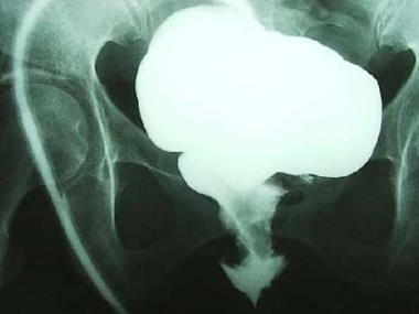 Cystogram of vesicovaginal fistula. Note the contrast extravasating from the bladder into the vaginal canal.
Other Tests
Methylene blue dye test
In patients who are suspected to have a vesicovaginal fistula, a methylene blue dye test can be performed, which has excellent specificity and is likely to be positive in diagnosis of fistula. Methylene blue is infused intravenously and a tampon is placed within the vagina. If the tampon turns blue, the test is positive for a vesicovaginal fistula.
Double dye test
Frequently, the double dye test is useful for diagnosing vesicovaginal and ureterovaginal fistulae. In this test, the patient ingests oral phenazopyridine (Pyridium), and indigo carmine or methylene blue is instilled into the bladder via a urethral catheter. Pyridium turns urine orange, and methylene blue (or indigo carmine) turns urine blue. A tampon is placed into the vagina. If the tampon turns blue, suspect vesicovaginal fistula. If the tampon turns orange, suspect ureterovaginal fistula. If the tampon turns blue and orange, suspect a combination of vesicovaginal and ureterovaginal fistulas.
Diagnostic Procedures
Cystoscopy
Cystoscopy with concurrent vaginal speculum examination helps determine the location and size of the fistula in relation to the vaginal cuff, trigone, and ureteral orifices. In addition, it reveals the degree of inflammatory reaction and the number of fistulas present. Most fistulas discovered after hysterectomy are located immediately behind the interureteric ridge and on the anterior vaginal vault.

Retrograde pyelography
This is the most definitive test to determine the presence of ureterovaginal fistula. Retrograde pyelography must be performed if IVP findings are abnormal or if the fistula site is difficult to locate. Performing bilateral retrograde ureteropyelography is often important because both ureters may be injured.
Histologic Findings
If the fistulous tract is excised as part of the repair technique, the specimen should be sent for pathologic evaluation to review the histologic findings. Pathologic findings vary depending on the cause of the fistula and may include foreign body, [13] giant cell reaction, malignancy, or chronic inflammation.
Giant cell reaction may be present if a foreign body was part of the cause of the fistula (eg, a nonabsorbable suture ligature of a uterine vessel catching the vaginal cuff and the bladder wall).
Radiation-induced fistulae are due to late changes caused by the radiation. After cessation of radiation therapy, fibrosis occurs in the bladder lamina propria. As fibrosis occurs in the subepithelial tissues, hyalinization of the connective tissues occurs. Often, large bizarre fibroblasts (ie, radiation fibroblasts) are encountered. An obliterative arteritis may be observed in medium-to-small vessels. These vascular changes may result in atrophy or necrosis of the bladder epithelium, causing ulceration or the formation of fissures. Again, it is important to rule out local cancer recurrence, especially in the setting of prior radiation therapy.
With fistulae due to cervical carcinoma, pathologic examination may demonstrate squamous cell carcinoma or adenocarcinoma. Fistulae due to iatrogenic injury manifest as signs of acute and chronic inflammation. The presence of abundant neutrophils suggests an acute inflammatory response. In patients with chronic inflammation, the predominantly lymphocytic infiltrate is associated with macrophages. In addition, interstitial tissue fibrosis and necrosis may be present.
-
Vaginal view of vesicovaginal fistula.
-
Cystoscopic view of vesicovaginal fistula.
-
Cystogram of vesicovaginal fistula. Note the contrast extravasating from the bladder into the vaginal canal.
-
This patient developed a supratrigonal vesicovaginal fistula immediately over the right ureteral orifice after transabdominal hysterectomy for uterine fibroids. The right ureteral orifice has been cannulated with a ureteral catheter to prevent injury to the ureteral orifice during the fistula repair. A Foley catheter has been inserted into the bladder. A transvaginal repair was performed.
-
Percutaneous suprapubic tube is placed prior to repair of a supratrigonal vesicovaginal fistula.
-
The supratrigonal vesicovaginal fistula site is marked out.
-
Supratrigonal vesicovaginal fistula. Isotonic sodium chloride is injected into the anterior vaginal wall to facilitate hydrodissection.
-
Supratrigonal vesicovaginal fistula. A J-shaped incision is made, and the anterior vaginal wall is dissected off proximally and distally to the fistula. The fistula site is not excised. A generous flap is created anteriorly and posteriorly to the fistula site. Surgical sutures have been placed in the fistula to close the site.
-
Supratrigonal vesicovaginal fistula. Surgical sutures are tied, and the fistula is closed.
-
Supratrigonal vesicovaginal fistula. Reinforcing tissue layers are used to cover up the fistula site in a nonoverlapping fashion. In this case, peritoneum followed by pubocervical fascia was used.
-
Supratrigonal vesicovaginal fistula. Vaginal wall is closed.


