Review

Carrot Anthocyanins Genetics and Genomics: Status and Perspectives to Improve Its Application for the Food Colorant Industry

1
Plants for Human Health Institute, North Carolina State University, Kannapolis, NC 28081, USA
2
Department of Horticultural Science, North Carolina State University, Raleigh, NC 27695, USA
3
Department of Horticulture, University of Wisconsin–Madison, Madison, WI 53706, USA
4
Vegetable Crops Research Unit, US Department of Agriculture–Agricultural Research Service, Madison, WI 53706, USA
5
National Scientific and Technical Research Council (CONICET), National Agricultural Technology Institute (INTA) E.E.A. La Consulta, Mendoza 5567, Argentina
6
Faculty of Agricultural Sciences, National University of Cuyo, Mendoza 5505, Argentina
*
Author to whom correspondence should be addressed.
Genes 2020, 11(8), 906; https://doi.org/10.3390/genes11080906
Submission received: 3 July 2020 / Revised: 31 July 2020 / Accepted: 31 July 2020 / Published: 7 August 2020

Abstract

:
Purple or black carrots (Daucus carota ssp. sativus var. atrorubens Alef) are characterized by their dark purple- to black-colored roots, owing their appearance to high anthocyanin concentrations. In recent years, there has been increasing interest in the use of black carrot anthocyanins as natural food dyes. Black carrot roots contain large quantities of mono-acylated anthocyanins, which impart a measure of heat-, light- and pH-stability, enhancing the color-stability of food products over their shelf-life. The genetic pathway controlling anthocyanin biosynthesis appears well conserved among land plants; however, different variants of anthocyanin-related genes between cultivars results in tissue-specific accumulations of purple pigments. Thus, broad genetic variations of anthocyanin profile, and tissue-specific distributions in carrot tissues and organs, can be observed, and the ratio of acylated to non-acylated anthocyanins varies significantly in the purple carrot germplasm. Additionally, anthocyanins synthesis can also be influenced by a wide range of external factors, such as abiotic stressors and/or chemical elicitors, directly affecting the anthocyanin yield and stability potential in food and beverage applications. In this study, we critically review and discuss the current knowledge on anthocyanin diversity, genetics and the molecular mechanisms controlling anthocyanin accumulation in carrots. We also provide a view of the current knowledge gaps and advancement needs as regards developing and applying innovative molecular tools to improve the yield, product performance and stability of carrot anthocyanin for use as a natural food colorant.

Graphical Abstract

1. Carrot as a Source of Anthocyanin for Natural Colorants

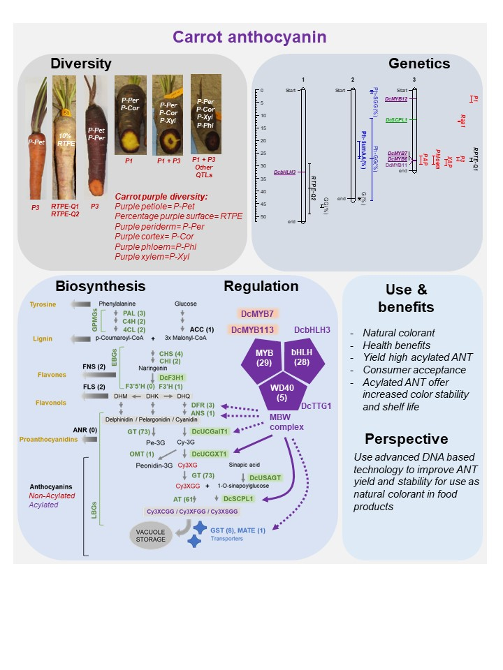
1.1. Application and Potential of Carrot as a Source of Natural Anthocyanins

The global market of natural colorants in the food industry was valued at USD 1.32 billion in 2015, and is expected to continue to grow over 7% annually between 2017 and 2022 [1]. This growth is due, in part, to the increased awareness of environmental hazards, the potential health benefits associated with the consumption of natural pigments, and the growing interest of the consumers in “natural” solutions [2]. To satisfy consumer demand, major food and beverage companies are moving towards replacing synthetic colors with plant-derived natural pigments in their products. However, the use of natural pigment is challenging due to interactions with food ingredients, the weaker tinctorial strength, the lower stability, and the challenges of matching the desired hues [3]. In addition, producing natural colorant is more expensive and requires efficient logistics. Indeed, while synthetic colorant is a more standardized industrial process, the production of natural colorants involves multiple steps, from development and production of the raw material, to extraction, dying and packing, which are more difficult to standardize. Comparisons of the synthetic vs. natural colorant prices are rarely reported in the scientific literature, but based on some food colorant industry experts, the cost of natural colors is about five times higher than synthetic colors on average, and in some cases (e.g., confectionery products) it can be as much as 20 times more expensive than synthetic (Peter Thorninger, https://www.nutritionaloutlook.com/view/switching-synthetic-natural-colors-here-are-your-challenges). Despite these challenges, the consumer demand for products that have a “clean label”, meaning they are free of artificial additives, continues to grow and represent the main driving factor for this shift [2,3]. For this reason, in order to achieve a sustainable natural colorant production system and market, it is becoming very critical to seek innovative solutions by taking advantage of the most advanced technologies.
Anthocyanin-rich extracts are currently among a group of commercially viable color ingredients, and are regulated as a color additive, although not defined as a “natural color” by the Food and Drug Administration (FDA). In the EU, water-based plant extracts containing anthocyanins within a natural range are widely permitted by the European Food Safety Authority (EFSA) for use as coloring in foodstuff due to their low toxicity [3], and are generally accepted as safe. However, when selectively extracted, further refined or concentrated, anthocyanins must be labeled with an E number (E163) as a color additive in the EU (Commission Directive 1333/2008 (EC).
Anthocyanins are a class of flavonoid antioxidants, and they represent some of the most vivid colors in nature, varying amongst shades of red, purple and blue, and they are produced in various plant tissues, including the leaves, roots, flowers and fruits. However, these natural pigments are generally unstable, and are susceptible to degradation driven by temperature, acidity and light exposure. In the past, this has limited their broad use as natural colorants. Recent advances in food technology and the identification of new sources of anthocyanins have contributed to a significant expansion of their use, especially in beverages. Anthocyanin’s share of the industry revenue has grown significantly over the last five-year period, reaching an estimated 9.5% share in 2018 [4]. Anthocyanins are likely to continue to be a key component of the “natural” food color industry, and remain a fast-growing category. The continued transition from artificial red, purple and blue color additives to anthocyanin-based pigments by food manufacturers will be driven by improvements in their chemical stability and versatility across broader food product forms.
In recent years, black or purple carrots have received much interest as natural sources of anthocyanins for application in the food industry (e.g., coloring soft drinks, fruit juices, jellies and confectioneries) [5]. Anthocyanin pigments extracted from purple carrot offer advantages over extraction from other fruit and vegetable sources due to the large concentration of anthocyanins in the former, which has been reported to be as much as 18 mg/100 g of fresh weight [6], and compared to other natural pigment sources (e.g., red cabbage) has low to no off-flavors. Additionally, a wide range of colors can be obtained from black carrot pigments, such as strawberry pink at low pH, versus purple and blue shades at higher pH [7,8]. This versatility is useful for coloring fruit juices, soft drinks and other food products [7], and could be used as a replacement for synthetic colorants such as FD&C Red 40 (allura red) [3,8,9]. Researchers investigating the color decay of ferric anthocyanin observed that during storage and thermal treatments, the pigment sources varied significantly; however, black carrot extract was the most promising pigment source of cobalt blue hues in pectin-stabilized solutions [10]. Compared to other anthocyanin-rich crops, like grapes, purple carrots contain lower amounts of non-anthocyanin phenolics that cause hazing and precipitation, and therefore carrots are a better source of anthocyanins [7]. Further, in a recent survey, 8 out of 10 consumers preferred seeing carrot extract on the food label to seeing synthetic colorant [11]. A review by Cortez et al. [12] highlighted that in the last five years, several processes for improving natural colorant stability using carrot extracts have been patented. New cultivars and methods for increasing anthocyanin production from carrot plants have also been developed and patented [13,14].
Acylated anthocyanins are an economically important colorant used in the food industry due to their increased stability at higher pH and their longer shelf-life [15]. In this regard, purple carrots also accumulate a high percentage of mono-acylated forms of anthocyanins [15,16], and unlike radish and red cabbage, the former do not require the removal of the sulfur aroma [15]. Acylation of anthocyanins is catalyzed by an acyltransferase [17], and acylation influences the cellular transport and stability of anthocyanins, which directly impacts their production, conservation, color, and thermal and shelf-life stability [15] when used as a natural dye or pigment [18,19]. Additionally, nonacylated anthocyanin are several times more bioavailable than acylated anthocyanin [20,21], and the consumption of carrot-derived anthocyanins may provide health benefits to the consumer [22]. For example, their high antioxidant capacity has been associated with protection against some forms of cancer [23,24], improved vision, reduced risk of diabetes [25], and enhanced cognitive and memory function [26]. Although there are no official statistics of worldwide purple carrot production, estimations by the food industry based on the annual need of 10,000 tons of carrot-derived anthocyanins place worldwide purple carrot production at approximately 100,000 ha [27].

1.2. Diversity for Anthocyanin Composition in the Purple Carrot Germplasm

The purple carrot germplasm exhibits broad genetic diversity, with regards to the total anthocyanin concentration and the distribution of these pigments across different root tissues. To date, several studies have characterized anthocyanin pigments—by HPLC analysis—in more than 30 carrot lines, including accessions from germplasm banks and open-pollinated (OP) and hybrid commercial varieties, reporting an overall range for total anthocyanin content of 0.5–191 mg/100 g fresh weight (fw) [6,28,29,30]. In purple carrot roots, the total anthocyanin content is strongly and positively correlated with total phenolics content, with correlation values (r) ranging from 0.85 [30] to 0.99 [31], suggesting that anthocyanins represent a large fraction of the phenolics in purple carrots. It is noteworthy that the purple color intensity and the extent of the root tissues covered with purple are both directly associated with the total root anthocyanin [32]. Figure 1 depicts the range of root phenotypic variation that can be found in the purple carrot germplasm. In most purple-rooted genetic backgrounds, anthocyanins are mainly expressed in the outer epidermal layer of the root, but can also be expressed in the cortex (outer-phloem), phloem and xylem (core) tissues, presenting different pigmentation patterns across these tissues, to the extreme extent of having all the root section fully saturated with the purple pigment (Figure 1). Thus, carrot accessions with dark purple color throughout the entire root section tend to have the highest anthocyanin content, whereas those with purple pigmentation in only the outermost tissues usually have low levels of anthocyanins. In most of the purple genotype evaluated to date, root surface is fully and uniformly purple. However, in a few studies [33], the color of the root surface was reported to be not uniformly purple, and the purple color was estimated as a percentage of the root surface [33]. Visual root analysis performed to date has indicated that purple germplasm from Syria (e.g., Homs type) and China (e.g., Ping Ding type) accumulate anthocyanin in the outermost epidermal cell layer, cortex and/or xylem, but are not fully purple. Carrot roots with purple in all their tissue layers are typically from Turkey. According to Bannoud et al. [30,34], when both phloem and xylem tissues were pigmented, the total anthocyanin content in the phloem was higher than in the xylem. In addition to root pigmentation, anthocyanins can accumulate in other tissues and organs of the carrot plant, including the bracts of inflorescences, the flower petals, the seed, the leaf petiole and lamina, and the nodes and internodes of the flower stalk [32].
Variation in root anthocyanin composition can be found in the carrot germplasm. Purple carrots have, predominantly, five cyanidin glycosides, two of which are non-acylated and three are mono-acylated anthocyanins (Table 1) [6,16,29,30]. However, in some studies, traces of pelargonidin and peonidin have been reported in some genetic backgrounds [6,29,36]. Among the cyanidin glycosides, five major compounds, two non-acylated and three acylated, are commonly found in purple carrots (Table 1). The percentage of acylated anthocyanins (AA) relative to the total anthocyanin content found across different studies varied from 25% to 99% [6,23,28,29,33], although in most commercial cultivars, AA predominate over non-acylated anthocyanins (NAA), with the former generally representing more than 60% of the total anthocyanins. In terms of absolute concentration, up to 155 mg/100 g fw of AA and ~36 mg/100 g fw of NAA have been reported in purple carrot lines [16].
Anthocyanin profiles vary across carrot genetic backgrounds. The most abundant root pigments are cyanidin glycosides acylated with ferulic (Cy3XFGG), sinapic (Cy3XSGG) and coumaric acid (Cy3XCGG), with Cy3XFGG being the main pigment in most of the accessions evaluated to date [16,28]. Coincidently with these reports, anthocyanin profiles in 208 purple-rooted carrots from an F2 mapping population generally revealed Cy3XFGG as the predominant pigment, followed by Cy3XSGG, representing on average 49% and 23% of the total anthocyanin content, respectively [33]. Similar results were found recently in an F2 population used for mapping QTL as conditioning root tissue-specific anthocyanins, with Cy3XFGG being the major pigment in both the phloem (accounting for 31% of total anthocyanins) and xylem (~52% of total anthocyanins), followed by Cy3XSGG, accounting for 23% and 34% of the total anthocyanins in these respective tissues, respectively. Among the non-acylated pigments, Cy3XG is generally found at a higher concentration than Cy3XGG, as observed in most commercial cultivars and accessions from germplasm banks [6,16,29,30], as well as in F2 and F4 populations recently characterized [34]. The opposite trend (i.e., Cy3XGG > Cy3XG) was found in another F2 previously described [33].
Characterizing the extent of the genetic and compositional variation in purple carrots is important from a breeding point of view, for both the production of chemically stable food dyes (e.g., aiming at developing cultivars with high concentrations of AA) and for the fresh market. Although purple germplasms have been previously characterized with regards to their anthocyanin composition, little information has been published to date on the evaluation of genetic diversity in these materials. In an early study, a large dataset of 4000 SNP markers was used to investigate carrot structure and domestication in a collection of 84 cultivated and wild carrot accessions from diverse geographical origins, including 9 purple-rooted accessions from Middle East and Central Asia [37]. The study revealed that all the purple carrots included in the study were genetically distinct, and they clustered with Eastern-cultivated carrots of other colors, clearly separating them from Western-cultivated and wild carrots from various origins [37]. More recently, Ipek, et al. [38] used 20 SSR markers to estimate genetic diversity in purple carrot populations of Ereğli, Turkey, composed of open-pollinated local cultivars and landraces. Substantial molecular variation in the SSR loci was found among these germplasms (i.e., 18 polymorphic SSRs yielded 106 alleles, and polymorphism information content (PIC) ranged from 0.29 to 0.85, with the expected heterozygosity ranging from 0.32 to 0.87), indicating a broad genetic diversity in these Turkish carrot populations. Although no pigment characterization was reported for these materials, these genetic resources are potentially valuable for the development of new purple carrot cultivars. Additional studies in other purple carrot germplasms, which combine both pigment compositional and molecular marker analyses, are necessary for a more detailed characterization of these materials, and to understand the origins of the purple root phenotypes from different genetic backgrounds. From a breeding perspective, the characterization of the purple carrot germplasms collected, based on their root anthocyanin content and AA:NAA ratio, will allow the selection of materials with high anthocyanin concentrations and specific pigment profiles, for the development of new varieties for either fresh consumption or the extraction of food colorants.

1.3. Shelf-Life Stability of Anthocyanins Derived from Carrot (pH, Temperature and Shelf-Life)

The shelf-life of naturally pigmented food products is, in general, reduced as compared to that of synthetic colorants, which results in expired and wasted food products. Variation in pH, temperature and light, as well as complexing with other compounds, can influence the stability of natural anthocyanin pigments during storage, and result in a modification or fading of the food color [39,40]. Acylated anthocyanins, as opposed to non-acylated anthocyanins, are the predominant anthocyanin-based colorants used by the food industry due to their greater color stability at a higher pH [15]. Acylation influences the cellular transport and stability of anthocyanins, directly influencing their accumulation and shelf-life conservation, when used as a natural dye or pigment [18,19]. Research into the stability of black carrot anthocyanins, regarding pH, temperature and shelf-life, has been thoroughly investigated [41]. Here we provide a summary of these studies, with an emphasis on the effect of the anthocyanin structure and carrot chemical composition on anthocyanin stability (Table 2). Storage at lower temperatures resulted in slower anthocyanin degradation as compared to storage at higher temperatures [42,43,44,45,46,47]. No significant effects on anthocyanin degradation were detected at temperatures below 4 °C and/or subfreezing [48,49]. At all temperatures, the NAA were significantly less stable than the AA [42,46,47]. For example, after storage at 40 °C, Turker et al. [42] reported that AA retained between 7.9% and 48.9% of their initial level, whereas NAA retained only 0% to 11% of their initial level. Compared to blackberry and acai anthocyanins, the rate of degradation of carrot anthocyanin has been shown to be slower [48].
The effect of pH has also been investigated in multiple studies [6,8,44]. In general, degradation rates of anthocyanin increased with pH, especially above 5. Montilla et al. [6] observed that Cy3XSGG exhibited a lower visual detection threshold at pH 3–5 than Cy3FGG and Cy3XCGG, suggesting that Cy3XSGG was more stable. In addition, a higher solid content was associated with a higher degradation of anthocyanins during thermal treatment (heating).
Comparing results from multiple studies, Kirca et al. [44] observed that anthocyanins from black carrot have greater stability during storage than those from blood orange, sour cherry, red-fleshed potato, red radish and blackcurrant during storage. Altogether, studies suggest that AA were significantly more stable than NAA at all storage temperature and pH ranges evaluated [41,42,46,50,51]. Furthermore, the high content of AA present in some black carrot genotypes has been suggested to be the key factor in explaining the higher stability of carrot anthocyanin as compared to other natural sources of anthocyanin.

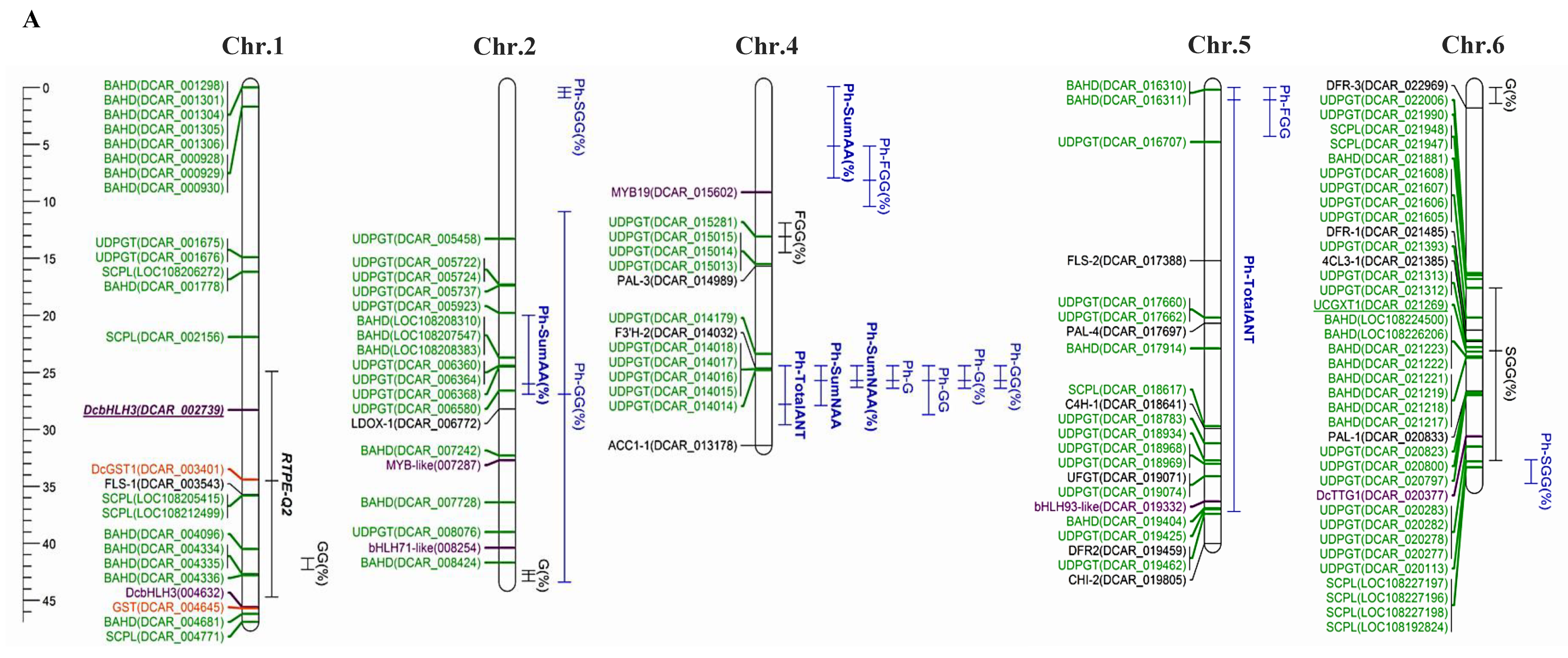
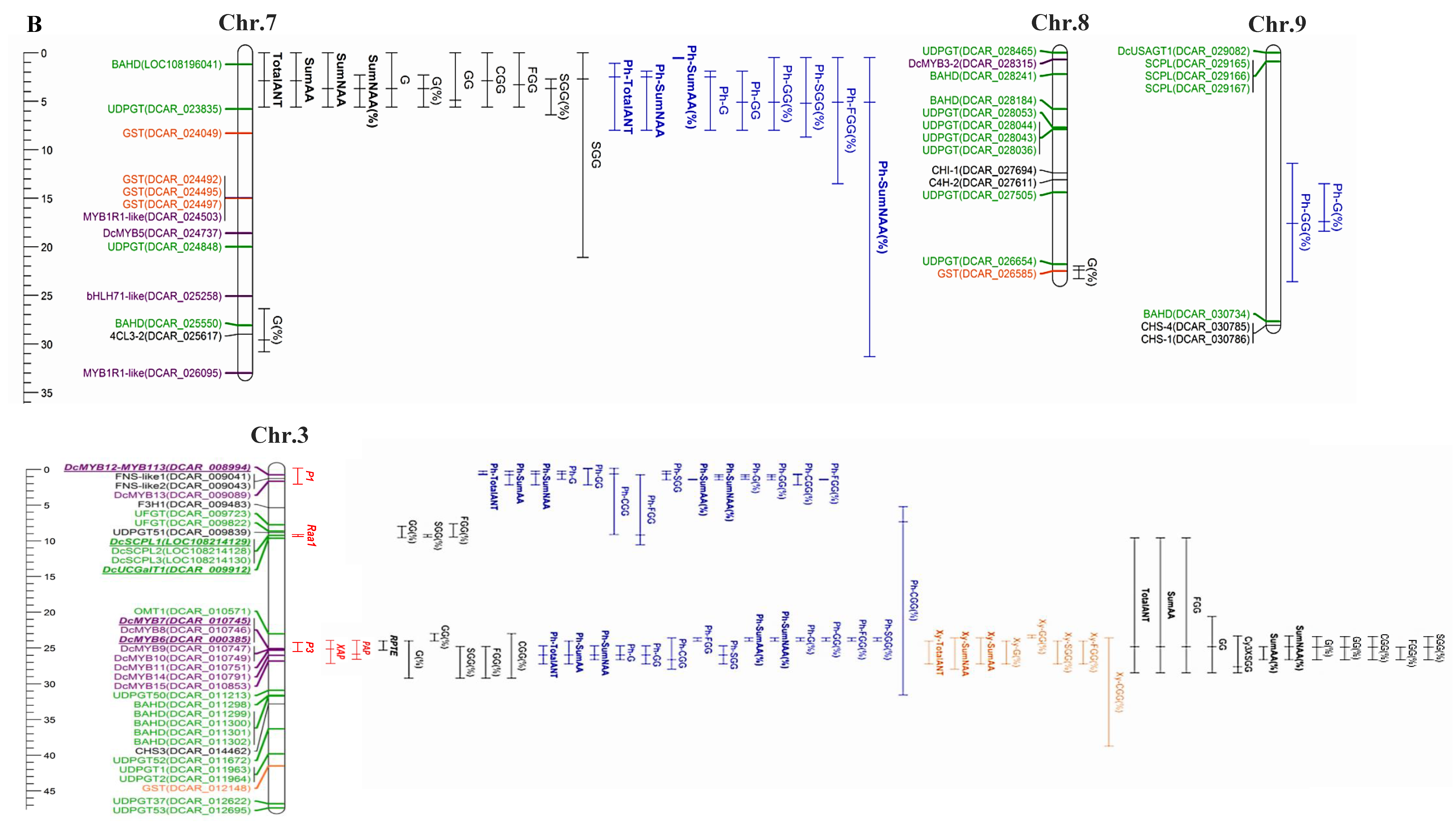
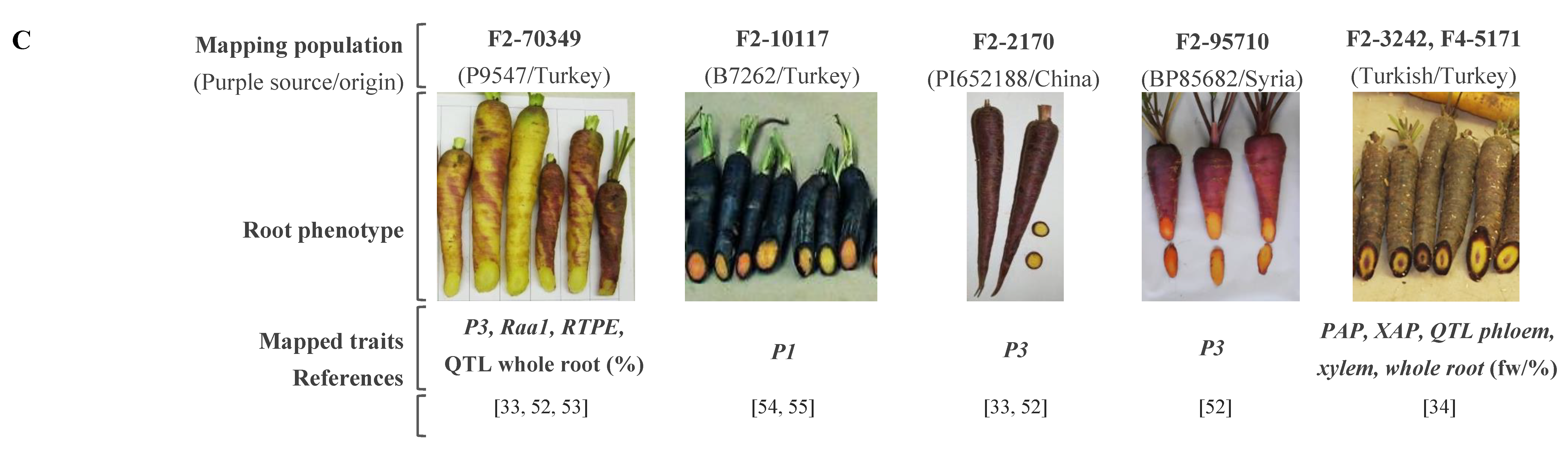
2. Genetics and Genes Controlling Anthocyanin Pigmentation in Carrot

2.1. Anthocyanin Genetics in Carrot

Important advances in the understanding of the genetic control of carrot anthocyanin pigmentation have been made. A summary of all mapped anthocyanin phenotypes and QTLs is reported in Supplementary Tables S1–S3, and is illustrated in Figure 2. In total, 158 loci or QTLs associated with anthocyanin accumulation in root or petioles were mapped for carrots. Among the different anthocyanidins detected in carrots, QTLs analyses were performed only for the cyanidin derivatives, since poenidin and perlargonidin derivatives were detected at a very low concentration.
A first study by Simon [57] described that a simply-inherited locus, called P1, controlled purple pigmentation in carrot roots, whereas another major locus (P2) conditioned pigmentation in the nodes, with P1 and P2 being genetically linked at ~36 cM. Subsequent studies were done by Vivek and Simon [54], and Yildiz et al. [55], into the Turkish ‘B7262’ genetic background, which presents purple pigmentation only in the outer root tissues (periderm and outer phloem) and has green petioles. Another dominant locus, called P3, conditioning anthocyanin accumulation in the root periderm and petioles of a Turkish (P9547) and a Chinese (PI652188) carrot lines, was later described and mapped to chromosome 3 [33]. Comparative analysis, segregating families derived from the B7262, P9547 and PI652188 backgrounds, revealed that P1 and P3 correspond to different loci in chromosome 3, and that the two loci are more than 30 cM apart [33].
In subsequent studies, segregation for root and petiole pigmentation was investigated and mapped to different genetic backgrounds, including an F2 developed from a Syrian purple carrot (BP85682), and advanced generations (F3, F5) of the mapping populations used previously by Cavagnaro et al. [33] and Iorizzo et al. [52]. Purple pigmentation in the root periderm and leaf petioles was fully co-segregated, and this suggested a single dominant gene for the genetic control of both traits. Comparative linkage analysis, with other populations harboring previously reported loci conditioning anthocyanin pigmentation, demonstrated P3-conditioned purple pigmentation in the Syrian background BP85682 as well. In another population, namely 5723, purple pigmentation in the petioles also segregated according to a 3:1 ratio, consistent with a single gene model, and this trait was mapped into the same region of P3. Thus, in some genetic backgrounds, P3 controls both root and petiole anthocyanin pigmentation, whereas in other backgrounds, purple petiole is independent of purple root. In B7262, ‘root pigmentation’ is conditioned by the P1 locus. This result suggested that within the P3 region, the loci controlling purple petioles and roots are tightly linked, but can act independently. Further, based on the extensive expansion of knowledge achieved in recent years regarding anthocyanin genetics in carrots, we here hypothesize that the control of the purple node, which was originally ascribed to P2 by Simon [57], actually corresponds to the P3 locus, which controls the purple petiole as mapped in the 5723 population. In addition to P1, P2 and P3, a simply-inherited trait, called Raa1 for ‘root anthocyanin acylation’, conditioning the percentage (%) of AA versus NAA, that is, the high % of AA being dominant over low % AA, was described and located—by linkage mapping—in chromosome 3, with its position being 17.9 cM from P3 [33]. Furthermore, very recently, Bannoud et al. [34] described and mapped two simply-inherited loci controlling the presence/absence of purple pigmentation in the root xylem and phloem, with purple being dominant over non-purple. These loci, called XAP and Phloem, for ‘xylem and phloem anthocyanin pigmentation’, were mapped in the same chromosome region of P3, together with another major locus controlling the presence/absence of pigmentation in the petioles, named PAP for ‘petiole anthocyanin pigmentation’ [34].
In addition to the simply-inherited loci controlling the presence or absence of anthocyanins in different root and leaf tissues, several quantitative trait loci (QTL) conditioning the concentration of root anthocyanins (Cy3XG, Cy3XGG, Cy3XFGG, Cy3XSGG and Cy3XCGG) have been described and mapped in the last six years [33,34,53,58]. In a first study, Cavagnaro et al. [33] mapped 15 QTL controlling the concentrations of four individual anthocyanin pigments (Cy3XG, Cy3XGG, Cy3XFGG and Cy3XSGG) as well as the total root anthocyanins (‘RTPE’, for ‘root total pigment estimate’) in an F2 family, named 70349, developed from the Turkish purple root source P9547. Segregation analysis for purple vs. non-purple root in 70349 and its F3 derivative populations indicated that two dominant loci interact epistatically in the genetic control of purple root pigmentation. The 15 QTLs were mapped to chromosomes 1, 2, 3, 6 and 8, and eight of them with the largest effects (26.6–73.3%) were co-localized to two regions of Chromosome 3. In the P3 region, the co-localization of a major QTL for RTPE (RTPE-Q1), which explained 50.5% of the variation, and QTLs for four root anthocyanins were found. A second QTL for RTPE (RTPE-Q2) explaining ~5% of the variation with lower phenotypic effect (~5%) was identified in Chromosome 1 (Figure 2). RTPE-Q1 and -Q2 explained the two-gene model observed for root purple color segregation in F2–F3 families. However, QTL interaction analysis indicated that RTPE-Q1 has a dominant effect, and is required for the expression of the RTPE phenotype. These results confirmed that the P3 region, where RTPE-Q1 was mapped, plays a key role in the expression of anthocyanin in carrot roots and petioles, and highlighted that a second QTL region (RTPE-Q2) mapped in chromosme 1 also influences—to a lesser extent—the total anthocyanin concentration in the carrot root.
In a recent study by Iorizzo et al. [52], high resolution mapping was performed for P3 using a larger population size of the same genetic background used by Cavagnaro et al. [33] (N = 187), reporting the identification and mapping of the same major QTL (Figure 2). A substantially smaller map region was attained for RTPE-Q1 and other root anthocyanin QTLs in this new map (Supplementary Table S2), as a consequence of the higher map resolution. Thus, in this region, Cavagnaro et al. [33] reported five overlapping QTL within a 12 cM region, whereas in the new map they spanned 6.3 cM, with co-localized QTLs for RTPE and three anthocyanin pigments within a 3 cM region.
In the study by Cavagnaro et al. [33], co-localized QTLs for the root AA Cy3XSGG and Cy3XFGG, and the NAA Cy3XGG, were also found in a small map region (3.6 cM) of Chromosome 3, and they all co-localized with Raa1. The QTL for Cy3XGG, which is proposed as the most likely substrate for acylation, had the highest LOD value (104.7), the largest phenotypic effect (73.3%) and the shortest confidence interval (0.7 cM) of all the 15 mapped QTL. These data suggest that Raa1 controls the ‘high’ versus ‘low’ percentage of acylated anthocyanins in carrot roots. Because the acylation of anthocyanins influences bioavailability [20,21] and pigment stability [59,60] understanding the genetic basis of anthocyanin acylation may be important for carrot breeding programs aimed at developing new cultivars with high levels of chemically-stable acylated pigments. Very recently, the Raa1 locus was characterized in detail by Curaba et al. [53] (described in Section 2.2).
In a newer study, Bannoud et al. [34] used two mapping populations (3242 and 5171) to map QTLs associated with total anthocyanin content, individual anthocyanin (Cy3XG, Cy3XGG, Cy3XFGG, Cy3XSGG and Cy3XCGG) content and relative percentages of individual anthocyanin, in the root phloem and xylem. In these two populations, anthocyanin accumulations in the phloem and outer-phloem (cortex) were not always distinguishable, and the purple pigmentation in these tissue layers was scored as phloem-specific (Cavagnaro personal communication). In total, 150 QTLs across seven chromosomes were mapped, with 8 of these QTLs associated with anthocyanin accumulation in the xylem, and 95 of the QTLs were mapped in chromosome 3 (Supplementary Tables S1–S3). Out of these 95 QTLs, 52 overlapping with the P1 region were associated with anthocyanin accumulation in the phloem, and 43 overlapping with the P3 region were associated with anthocyanin accumulation in the phloem and xylem. The other 24 and 12 QTLs associated with anthocyanin accumulation in the phloem mapped to two overlapping regions on chromosome 4 and 7, respectively.
Overall, across all genetic studies for anthocyanin pigmentation in carrots, three QTL regions (P1, P3 and RTPE-Q-2) control the presence of purple pigmentation in the carrot root in a tissue-specific manner, one QTL region controls purple pigmentation in the petiole (P3), and another QTL region (Raa1) controls anthocyanin acylation. Across all studies, the P3 region has been identified as a candidate region harboring the key gene(s) controlling anthocyanin accumulation across all tissues, while the P1 region is involved in the regulation of anthocyanin accumulation in the root out-phloem and/or phloem of specific genetic backgrounds. Several other QTLs with lower effects were identified, and will serve as a foundation for studying the overall molecular mechanisms and their interactions controlling anthocyanin synthesis, storage and degradation. Given the tissue-specific nature of anthocyanin accumulation in carrot, by dissecting the genes involved in tissue-specific anthocyanin expression, breeders will gain a better understanding of the genetics underlying these traits, and may be able to predict root color phenotypes in directed crosses. DNA markers associated with anthocyanin accumulation in carrots have been identified, and the regulatory and structural genes involved in the anthocyanin biosynthetic pathway are being investigated [56,58].

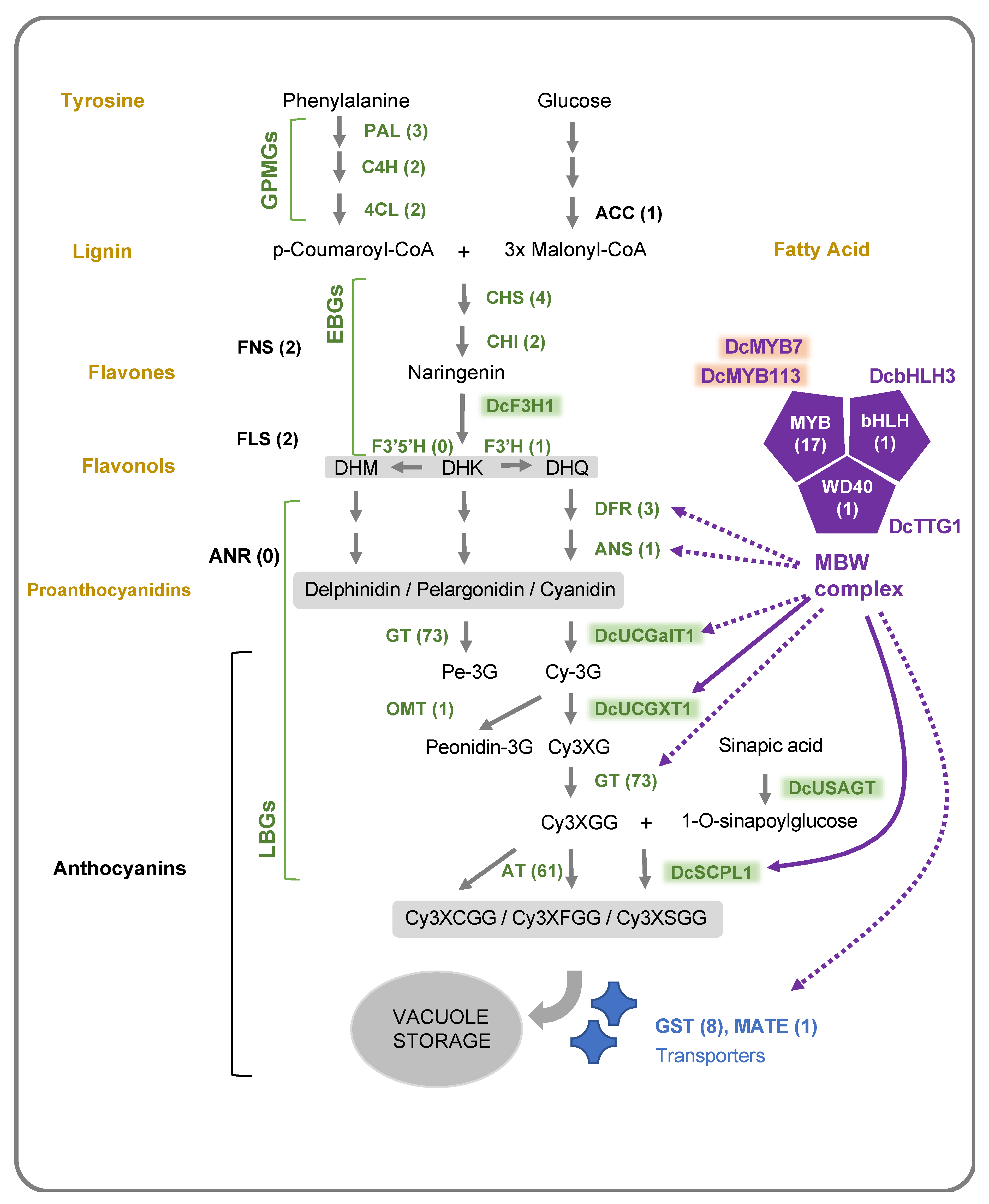
2.2. Anthocyanin Structural Genes

Anthocyanin accumulation is determined by the activity of structural genes, which are divided into general phenylpropanoid metabolism genes (abbreviated here GPMGs) and early and late biosynthesis genes (EBGs and LBGs, respectively) [61,62]) (Figure 3). GPMGs are required for the synthesis of other phenylpropanoids, such as lignin and Acetyl-CoA carboxylase (ACC), for the production of fatty acid compounds containing 4-coumaroyl-CoA and malonyl-CoA, respectively [61,63]. EBGs are shared for the biosynthesis of multiple flavonoids, whereas LBGs are more specific to the anthocyanins [63,64]. LBGs include genes coding for modification enzymes, such as glycosyltransferases (GTs) and acyltransferases (ATs), which catalyze the addition of sugar moieties and acyl groups, respectively, resulting in specific decoration patterns that greatly influence their function and stability. Since the carrot genome was released in 2016, curated annotation identified 159 potential structural anthocyanin genes, either located within an anthocyanin-related QTL or differentially expressed between purple and non-purple tissues [34,53,56,65,66,67] (Supplementary Table S4). These include 8 GPMGs, 8 EBGs and 139 LBGs, including 73 GTs genes, 61 ATs genes and 1 O-methyltransferase (OMT) gene, coding for enzymes involved in anthocyanin glycosylation, acylation and methylation, respectively (Supplementary Table S4). Comparative analysis with other genomes, like grapevine and Arabidopsis, indicated that the carrot genome lacks the Flavonoid 3’5’ hydroxylase (F3′5′H) and the anthocyanidin reductase (ANR) genes. The ANR enzyme catalyzes the first step of the proanthocyanindin (PA) pathway (Figure 3), and F3′5′H is required to direct the flux toward the anthocyanin delphinidin derivatives, which perhaps partially explains the low diversity of the anthocyanin and flavonoid derivatives detected in carrots. The characterization of genes retained after three whole genome duplications (WGD) indicated that several flavonoid/anthocyanin genes are duplicated. For example, three copies of the phenylalanine ammonia-lyase (DcPAL1, DcPAL3 and DcPAL4) were retained after each of the three WGDs [56]. Several ATs and GTs were organized in tandem clusters [53], likely as a result of recent tandem duplications [68,69]. Although the role of these duplicated genes in the carrot is still unknown, each of these duplicated genes may have acquired a specialized function in the expression of the pathway in specific tissues, or under specific environmental conditions (e.g., abiotic stresses).
The expression data from eight independent studies are available for 105 anthocyanin structural genes, including 90 with a detectable level of mRNA above a 1 RPKM threshold [34,52,53,55,67,70,71,72] (Supplementary Table S4). In this review, we integrated all these data so as to highlight differences and consistency. Differential gene expression analysis of purple vs. non-purple tissues identified 78 genes as being up or downregulated in at least one genotype, 54 of which were found to be upregulated in at least one purple root sample, including 7 genes (DcPAL4, DcC4H1, DcCHS1, DcCHI1, DcF3H1, DcF3’H1 and DcDFR1) consistently reported to be upregulated in at least nine independent carrot lines (Table 3). This may indicate that the transcriptional regulation of these seven genes was targeted early in the evolution of purple carrots, and may be well conserved among the various carrot cultivars used in breeding today.
The expression level in the petiole, which was only measured in one study, revealed that five glycosyltransferases coding genes (DcUDPGT1, 8, 32, 50 and 70) and DcFLS2 could be specifically active in the aerial parts [52] (Supplementary Table S4). The downregulation of DcFLS2 in purple pigmented petiole, associated with the upregulation of DcDFR1, could significantly contribute to directing the metabolic flux of dihydroflavonols toward the anthocyanin pathway in this tissue, as it was observed in the corolla of other plant species [73,74]. The highest level of anthocyanin accumulation could be achieved when both genes were antagonistically regulated [75]. Similarly, two flavone synthase like genes (DcFNS-like1 and DcFNS-like2) and two flavanone hydroxylases (DcF3H and DcF3′H), which compete for Naringenin as substrate, present opposite expression patterns in dark vs. pale purple phloem [34]. Increasing the level of FNS in transgenic celery dramatically reduces the anthocyanin content, as well as the expression levels of F3′H and DFR, suggesting the existence of a molecular mechanism coordinating their expression in the Apiaceae family [76]. Reducing FNS activity in black-colored dahlia plants was also linked to the accumulation of high amounts of anthocyanins [77]. Interestingly, however, a minimum level of FNS activity is needed to produce flavone co-pigments which could help in stabilizing the accumulation of anthocyanins [78]. The transcriptional regulation of genes coding for metabolic branching point enzymes, such as FNS/F3H and FLS/DFR, plays a critical role in balancing the metabolic flux of phenylpropanoids, and is likely to be a determinant factor in the production of purple pigments in carrot [74,79,80,81]. Among all the structural genes tested, DcDFR1 is the only one that is always found upregulated in all purple tissue, making it the most reliable marker for anthocyanin biosynthesis and a possible bottleneck of the pathway in carrot. Indeed, increases in DcDFR1 expression tend to be proportional to the accumulation levels of anthocyanins observed in a carrot population that segregated for purple color intensity in the root phloem [34].
Among the annotated carrot anthocyanin structural genes, five were functionally characterized: DcF3H1 [83], DcUCGalT1 [65], DcUCGXT1 [72], DcSCPL1 [72] and DcUSAGT [66] (Figure 3, Table 3 and Supplementary Table S4). The knockout of DcF3H1 (DCAR_009483) using the CRISP/Cas9 system caused the discoloration of calli, which validated the function of this gene in the biosynthesis of anthocyanin in carrot, as well as demonstrating the successful application of CRISPR/Cas9 in carrots. DcUCGalT1 (DCAR_009912) has been shown to catalyze the formation of cyanidin 3-galactoside (Cy3G) in vitro, and DcUSAGT (DCAR_029082) was shown to catalyze the transfer of a glucose moiety to the carboxyl group of sinapic acid, thereby forming 1-O-sinapoylglucose [65,66]. 1-O-sinapoylglucose serves as an acyl donor in the acylation of cyanidin-3-(2″-xylose-6-glucose-galactoside) (Cy3XGG) into its acylated counterpart, cyanidin-3-(2″-xylose-6″-sinapoyl-glucose-galactoside) (Cy3XSGG), which helps stabilize the accumulation of anthocyanins in purple carrots. This reaction was recently attributed to DcSCPL1 (also named DcSAT1) by two independent studies [53,72]. DcSCPL1 was first identified as a strong candidate for the Root Anthocyanin Acylation 1 (Raa1) locus controlling the formation of Cy3XSGG and Cy3XFGG in the carrot storage roots of three mapping populations [53]. Sequence analysis of DcSCPL1, in both high and low acylated backgrounds, revealed the presence of two distinct alleles, one functional and the other not, that can easily be identified by PCR. Another study by Xu et al. [72] supported this hypothesis by showing that overexpressing DcSCPL1 in the calli of the dark purple-rooted carrot ‘Deep purple (DPP)’ increases the production of Cy3XSGG [72].
In this review, we localized the physical location and boundaries of all published QTLs, and annotated the anthocyanin structural genes identified in carrots to date, noting that multiple LBGs and EBGs were localized within anthocyanin QTLs (Table 3; Supplementary Table S4). This includes, for example, a cluster of four DcSCPL-ATs (LOC108227197, LOC108227196, LOC108227198 and LOC108192824), of which three are upregulated in purple roots, and are co-localized with two QTLs for Cy3XSGG, mapped in chromosome 6 [34]. Interestingly, Curaba et al. [53] noted that two of these genes, LOC108192824 and LOC108227198, clustered with DcSCPL1 in clade IA-1, and possess the predicted functional SCPL domain/motifs. One BADH-AT (LOC108196041) co-localized with 15 QTLs on chromosome 7, of which 9 were QTLs associated with a potential acylation function (Table 3; Supplementary Table S4). Furthermore, a cluster of 5 LBG-GTs and DcF3’H1 overlapped with 12 phloem-specific QTLs located in chromosome 4, which include a QTL for total anthocyanin [34]. Interestingly, in multiple studies, DcF3′H1 is found to be upregulated in purple carrot roots, and is highly co-expressed with DcMYB113 [72], a transcription factor controlling anthocyanin accumulation in the periderm and phloem (considered as outer-phloem in this review) (Supplementary Table S4). This is the first time that these published results have been integrated, and therefore these new findings provide new opportunities to further investigate the genetic pathway controlling the synthesis and various decoration patterns of anthocyanins present in carrots, which likely involves multiple AT and GT genes coding for specialized enzymes with different substrate specificities.
Overall, across all the published anthocyanin genetic studies in carrots, none of the chromosome locations harboring GPMGs or EBGs overlapped with any of the major QTL regions (P1, P3, RTPE-Q-2, XAP, PAP or Phloem) controlling the expression of anthocyanins in purple root tissues and petioles. The expression levels of most structural genes related to flavonoid metabolic pathways are consistently higher in purple vs. non-purple tissues. Duplicated genes appear to have divergent expressions of root tissue-specificity, such as DcCHS1, which is upregulated mostly in purple xylem, compared to DcCHS2 and DcCHS9, which are upregulated mostly in purple phloem tissues, or DcDFR1, which is upregulated in purple xylem as opposed to DcDFR2 and 3, two genes that are slightly downregulated in the same tissue [67] (Supplementary Table S4). These results suggest that the purple phenotype is likely controlled by more than one of the transcription factors that control anthocyanin accumulation in carrot root and petioles by coordinating the expression of structural genes in a tissue-specific manner.

2.3. Regulatory Anthocyanin Genes

The regulation of the expression of anthocyanin structural genes, especially LBGs, is coordinated by the MYB-bHLH-WD40 (MBW) protein complex, in which the role of the MYB and bHLH transcription factors is critical to triggering anthocyanin accumulation in specific tissues [62,63,84,85,86,87] (Figure 3). The regulation of LBG activity by the MBW complex is well conserved among land plants, and several studies report the functional characterization of MBW members through transgenic expression in orthologous system [61,62,72,88,89,90,91,92,93,94]. Among 891 MYB-, bHLH- and WD40-coding genes mapped in the carrot genome by Iorizzo et al. (2019), 73 genes are potentially related to the anthocyanin metabolism through one of the following three criteria: overlapping with an anthocyanin-related QTL, differentially expressed between purple and non-purple tissues, or orthologous to a known anthocyanin-related gene from another species (Supplementary Table S4) [34,52,67,71,72,95]. Only 12 of them, including 11 anthocyanin-related MYBs (A-MYBs) and 1 anthocyanin-related bHLH (A-bHLH), DcbHLH3, possessed all three criteria, and therefore represent primary candidates for further investigating the regulation of purple pigmentation in carrot (Table 3). RNAseq data from specific tissue layers offers valuable information in order to identify the candidate genes controlling anthocyanin accumulation in the storage root. However, to date only a few studies have performed comparative transcriptome analysis by sampling specific root tissues. This is due in part to the limited knowledge that the carrot breeding and genetic community has regarding the genetic inheritance of anthocyanin accumulation in the different tissues, and also to an often inconsistent identification/naming of the different purple root tissues sampled/analyzed across studies. Considering that the most striking differences in purple pigmentation between carrot cultivars are between xylem and phloem tissues, it is interesting to notice that 10 A-MYBs and 14 A-bHLHs, including bHLH3, present a differential regulation between these two tissues (Supplementary Table S4). A-MYBs in other plant species were identified as either activators or repressors of the anthocyanin pathway [86,96,97]. Transcriptome data of carrots revealed that 4 A-MYBs, including 2 that were predicted as MYB1R1-like transcription factors (DcMYB1R1-1 and DcMYB1R1-2), were downregulated in both phloem and xylem tissues, and may negatively regulate anthocyanin biosynthesis [67] (Table 3; Supplementary Table S4). Such repressors could inhibit the accumulation of anthocyanin by directly repressing the expression of structural genes, interfering with the MBW activity, or promoting the expression of competing enzymes that use the same substrates required for anthocyanin production [97]. Examples can be found in other plant species, such as in strawberries, wherein FaMYB1 can interact with bHLH proteins and repress the expression of structural genes at the lower end of the flavonoid pathway (ANS and GT) [94], or in the Mimulus lewisii flower, wherein LAR1 represses the biosynthesis of anthocyanin by activating the expression of FLS [73].
Using a fine mapping approach, Iorizzo et al. [52] identified a cluster of six genes coding for A-MYB transcription factors (DcMYB6, 7, 8, 9, 10 and 11) in the P3 region harboring overlapping QTLs (Table 3; Supplementary Table S4). The functional characterization of DcMYB6, using non-endogenous 35S promoter, demonstrated its ability to induce anthocyanin expression in Arabidopsis, but not in orange Kurodagosun (KRD) carrots [71,90]. The expression data indicate a genotype-specific regulatory activity for DcMYB6. While highly expressed in most purple root cultivars, DcMYB6 expression does not correlate with anthocyanin pigmentation in all the purple-rooted carrot lines tested, and remains expressed in some non-purple tissues [34,52,71]. Within the A-MYB cluster, DcMYB7 is the only gene specifically overexpressed in all purple vs. non-purple root tissues, and its expression was also detected in the purple petiole of two carrot lines [52,71] (Table 3; Supplementary Table S4). Further studies from the transgenic approach, using both overexpression and knockout carrot lines, showed that DcMYB7 is functional in at least three purple cultivars, and is essential for the production of purple pigments in at least one of them (DPP), making it the best candidate gene for the P3 locus controlling anthocyanin pigmentation in the carrot storage root [71,98]. DcMYB11 is the only A-MYB specifically expressed in all purple petiole, and represents the best candidate for the genetic control of petiole purple pigmentation (Table 3; Supplementary Table S4). More recently, another MYB transcription factor, DcMYB113, was identified as a candidate for P1, and was functionally characterized [72]. The expression of DcMYB113 appears to be cultivar-specific, and restricted to the root periderm and phloem of the carrot cultivar ‘purple haze’ (PPHZ) (Table 3; Supplementary Table S4).
By identifying the key A-MYB regulatory gene(s), it may be possible to initiate and control the entire anthocyanin pathway in carrots. Indeed, the overexpression of either DcMYB7 or DcMYB113 in orange KRD cultivar triggers the accumulation of anthocyanins in the entire carrot tap root and petiole, although only DcMYB7 under the control of a strong 35S-promoter was reported to induce purple pigmentation in the reproductive organs of the transgenic lines [71,72]. Both A-MYBs can interact with DcbHLH3, and could directly activate the expression of two structural genes related to anthocyanin structural modification—a glycosyltransferase, DcUCGXT1 and an acyltransferase, DcSCPL1 [71,72]. DcbHLH3 co-localize with the RTPE-Q2 in chromosome 1, one of two major QTLs controlling anthocyanin accumulation in a mapping population derived from a Turkish carrot used as the purple root source progenitor [33]. Interestingly, the overexpression of DcMYB113 in KRD leads to an increase in the Cy3FGG/Cy3SGG ratio, whereas the overexpression of DcMYB7 in the same cultivar may have the opposite effects [71,72]. The differential activity of A-MYBs could be responsible not only for variations in the levels of anthocyanins being produced, but also their profile.
Among the five annotated carrot anthocyanin-related WD40 (A-WD40) genes, only DcTTG1 transcripts have been detected in carrot roots so far [52,95]. DcTTG1 was identified by Kodama et al., on the basis of its homology via Blast analysis to Arabidopsis TTG1. Here, we confirmed by orthologous, phylogenetic and synteny analysis that DcTTG1 cluster with TTG1 (Table 3; Supplementary Table S4). TTG1 is constitutively expressed in all major organs, and is a constant member of the MBW complex required for the activation of the anthocyanin pathway and the determination of epidermal cell fate in Arabidopsis [99,100,101]. In carrots, DcTTG1 is located in chromosome 6, and its location does not overlap with any anthocyanin-related QTLs previously mapped in carrots (Table 3; Supplementary Table S4). Investigation into the transcriptome analysis results for this gene from Iorizzo et al. [52] and Curaba et al. [53] confirms that DcTTG1 is constitutively expressed, although a possible positive correlation between transcript abundance and total anthocyanin content was reported by Kodama et al. [95] in one cultivar. This suggests that DcTTG1 may exist only as a functional gene in carrots, and could play a central role in the formation of the MBW complex similar to that observed in Arabidopsis. Although these results are preliminary, they provide directions to further study the MBW protein complex in carrots, and its impact on the regulation of the anthocyanin biosynthetic pathway.
The regulation of anthocyanin metabolism ends with their transport into the vacuole, a process which involves Glutathione S-transferases (GSTs) [102,103]. DcGST1 (DCAR_003401), which co-localized with RTPE-Q2 QTL, was recently identified as being upregulated in two purple carrot cultivars [67], and its expression could be directly regulated by an A-MYB as it was found in Arabidopsis and apple plants [104,105]) (Table 3; Supplementary Table S4). In fact, based on the cultivar- and tissue-specific expression patterns, DcMYB113 likely controls the expression of DcGST1, and does this independently of DcMYB7 [71,72]. Interestingly, DcMYB113 is also co-expressed with DcMATE1 (DCAR_031151), another potential anthocyanin transporter [72,102]. A-MYBs are central to the regulation of anthocyanin biosynthesis genes, however the molecular mechanisms controlling their activity during carrot development remain largely unknown.

3. External Factors Affecting Anthocyanin Accumulation and Profile in Carrots and Other Plant Species

Phenolic compounds such as anthocyanins are essential to the interactions between plants and their environments [106]. Although some organs of some plant taxa can synthesize and accumulate anthocyanins in a nearly constitutive fashion (e.g., black carrots and red grapes), in other plants the accumulation of anthocyanins may reflect an adaptive response to adverse environmental conditions, and such accumulation of phenolics is considered an indicator of plant stress. Chemical elicitors or abiotic stresses could induce the accumulation of anthocyanin in carrots, and be an effective alternative and/or complement to breeding [107].
Various plant species show a similar anthocyanin induction in response to the same stresses, suggesting that the molecular mechanisms controlling anthocyanin stress responses are, at least partially, conserved among land plants. Temperature, light, nutrients and water intake are all environmental conditions that can affect anthocyanin biosynthesis [62,102,108]. Although not completely understood, their accumulation in response to abiotic stress has been associated with an increase in plant survival rate, which is likely due to their protective role against reactive oxygen species (ROS) [86,108,109]. Abiotic stress signaling was shown to affect the activity of several EBGs and LBGs genes in many plant species, coinciding with the production of anthocyanin. Such coordinated action likely involves the regulation of components of the MBW complex, and in particular A-MYBs and A-bHLHs [61,62,86,97,110,111].
Here we have summarized the effect of external factors on anthocyanin accumulation in carrots, and a few examples from some other species (Table 4). The treatment of carrot plants with exogenous phytohormones could also affect the production of anthocyanin. Indeed, foliar-applied ethephon, a precursor of ethylene (ET), enhances the content of anthocyanin and total phenolic compounds in Deep Purple carrot roots by about 25%, indicating that the production of anthocyanin can be increased even in black carrot varieties already containing a high level of anthocyanins [112]. In blueberries, apples and lettuce, the exogenous application of jasmonic acid (JA) increases the total phenolics content and antioxidant capacity [113,114,115]. In Arabidopsis, both abscisic acid (ABA) and JA promote the biosynthesis of anthocyanin in the presence of sucrose, while gibberellic acid (GA) and ET repress anthocyanin production [116,117,118,119]. The exogenous application of sucrose, which is perhaps the most potent inducer of anthocyanin biosynthesis, was reported in several plant species [120,121,122,123,124,125]. Although the effect of sucrose treatment in carrot has never been reported in planta, a significant increase of anthocyanin accumulation up to 7.5-fold was observed in carrot cell cultures [126,127] (Table 4). In Arabidopsis, the largest contributor of sucrose-induced anthocyanin accumulation was found to be PAP1, an ortholog of DcMYB6 and 7 [56,128]. Interestingly, the transcript level of one of its homologs, PAP2, was found to increase about 1000-fold in response to nitrogen deficiency, and was proposed to be the mean mediator of anthocyanin accumulation in response to this stress [129], indicating the specification of A-MYBs to different stress-response pathways. A-MYBs’ activity can also be stress-regulated at the post-transcriptional level, as shown by the ubiquitination and degradation of MdMYB1 in response to increased nitrogen intake in apple tissue cultures [130]. Coherently, nitrogen concentration was shown to affect the production of anthocyanin in carrot cell cultures, but conflicting results were obtained from two independent studies, with both positive and negative effects of nitrogen being observed [126,127]. Altering the source of nitrogen by modifying the balance of ammonium to nitrate significantly affects the production of anthocyanin in carrot cell cultures, with a 1:4 ratio being optimum [127]. Additionally, reducing phosphorous intake was shown to enhance the production of anthocyanin in both carrot cell culture and Arabidopsis [109,126].
The application of postharvest abiotic stresses to carrots, such as wounding, could promote the accumulation of purple pigments. The activity of DcPAL1, along with the expression of DcPAL1, DcC4H1 and Dc4CL3-1, significantly increases in wounded carrots, therefore promoting the metabolism of phenylpropanoids and the production of many of the phenolic defense compounds [133,134,135,140]. Wounding stress increases, by 75%, the total phenolic content in shredded carrots stored at 15 °C for 6 days [132]. Combining wounding with additional stresses such as heat, UV light, hyperoxia or the application of phytohormones and herbicide can synergistically increase the accumulation of phenolics in carrots [107,132,135,136,139,140,141]. For example, excess oxygen [107], higher storage temperatures [138], UV-B radiation [137] and ET [132] can enhance the content of total phenolics in wounded carrots 3.5-, 4.8-, 3.2- and 1.7-fold, respectively. Interestingly, ET and hyperoxia have little to no effect on non-wounded harvested carrots [132,135]. Although no detailed information exists concerning the anthocyanin profile in wounded carrots, the activation of the phenylpropanoid pathway and enhanced antioxidant capacity suggest that anthocyanins could be produced by wounding in some genetic backgrounds [132,134,135,137,138]. This correlates with the observation that combined light and mechanical stress can increase by, about 50%, the accumulation of anthocyanin in carrot cell cultures [142]. Additionally, the increases level of organic acids, such as ferulic acid, in response to wounding could improve anthocyanin stability through co-pigmentation effect [132,134,143].
Despite the observed effects of sugars, minerals and phytohormones on anthocyanin accumulation, to date no study has investigated the interaction between these external factors and anthocyanin-related genes in carrot. Extensive variations of anthocyanin-related TFs have been identified, and polymorphism within their promoter regions, such as was reported for DcMYB7 alleles, suggests that a range of sensitivity to stress-induced anthocyanin responses could be observed between carrot cultivars [71,128]. Additionally, as observed in other plant species, exposure to various abiotic stress conditions could differentially affect the regulation of structural genes, and therefore the composition and tissue localization of anthocyanins being produced [108,123,144,145,146]. Two UDP-GT enzymes contribute to cold, salt and drought stress tolerance via modulating anthocyanin accumulation in Arabidopsis [131]. Such an osmotic stress response can be induce by mannitol, which promotes anthocyanin accumulation in both Arabidopsis and carrots [126,131]. Understanding the mechanism controlling anthocyanin’s response to stress would help us in developing new strategies to maximize the use of carrot as a natural colorant. Combined stresses can have an additive effect on the anthocyanin accumulation, and evidence from Arabidopsis suggests that the regulation of anthocyanin biosynthesis integrates independent and reversible stress-induced pathways. For example, in Arabidopsis, the accumulation of anthocyanin in response to salt stress is controlled independently of other stresses, such as high light, low phosphate limitation, high temperature or drought [147], and nitrogen and phosphorus depletion were found to trigger anthocyanin production through distinct pathways [109]. Most abiotic stress studies in carrots are postharvest, and more research to understand their effects on anthocyanin-regulatory genes during the development of the storage root is needed. For instance, extended growth was shown to significantly increase the concentration of anthocyanin in the storage root of the ‘Deep Purple’cultivar [148].

4. Perspectives

4.1. Advancing Molecular and Biotechnology Tools to Develop Carrot Cultivars That Maximize Anthocyanin Yield in Product Performance and Stability

Classical plant breeding approaches have succeeded in improving the productivity and quality of carrots for producers and consumers over the last century [149]. A significant focus in breeding has been placed on breeding for male sterility, disease resistance, vernalization requirement, root morphology and carotenoid content, with limited interest in anthocyanin-related phenotype/profile. Given the growing interest in multi-colored carrots and their nutritional profile, a few purple carrot cultivars have been released over the last few years [13,150]. However, as carrot breeding programs move forward, the expansion of carrot global markets and a broader range of consumer traits will require attention, including those related to anthocyanin accumulation for use as a natural food colorants. To improve anthocyanin content, profile and stability in well-established classic carrot breeding strategies can be adopted, and future work will need to focus on expanding molecular tools to facilitate the incorporation of multiple phenotypes into new cultivars. Commercial breeders today use molecular markers to implement Marker-Assisted Breeding (MAB) as a strategy for the effective improvement of multiple traits in plants [151]. MAB includes marker-assisted selection (MAS), marker-assisted recurrent selection (MARS), marker-assisted backcrossing (MABC), and genome-wide selection (GWS) or genomic selection (GS) [151]. Establishing DNA marker assays that can effectively be used for MAB is critical in order to bridge the gap between researchers discovering new QTLs and gene-trait associations, and breeders using this knowledge to make informed breeding decisions. Besides MAB, emerging biotechnology applications, such as gene editing, promise opportunities to effectively integrate desired traits into new cultivars. The application of this biotech method demands a more detailed understanding of the molecular mechanisms controlling desirable traits.
As described in this review, the number of marker trait association studies concerning anthocyanin in carrots has increased considerably over the last few years, leading to the identification of multiple genes controlling major QTLs and simply-inherited traits. Despite these advances, very few DNA marker assays targeting anthocyanin or other traits have been developed for carrots, thus limiting the implementation of MAB in carrot breeding programs. For example, a cleaved amplified polymorphic sequences (CAPS) marker has been developed for the Y2 locus controlling carotenoid accumulation [152]. Similarly, a PCR-based marker, targeting the DcSCPL1 gene controlling anthocyanin expression associated with the Raa1 locus, is able to differentiate the low and high acylation alleles [53]. However, all of these markers were tested in just a few mapping populations, representing a very narrow genetic diversity. Therefore, it is unknown if these DNA assays would be successful in other genetic backgrounds. The reliability of molecular markers in predicting the target trait depends on their close linkage with the mutation that effects the phenotype [153]. This is particularly important in outcrossing species, such as carrots, in which Linkage Disequilibrium (LD) decays rapidly [152], and identifying causal mutations that are present in perfect LD with the phenotype is critical for effective MAB application. Considering the high genetic variation existing in the carrot germplasm [152], it is common that PCR primers developed for one mapping population do not work in other genetic backgrounds. In order to implement MAB, these QTLs need to be validated in wider breeding populations and germplasm collections, and under different growing conditions. However, all of these PCR-based marker methods are low throughput, relatively expensive, and are labor intensive, which limits their use in carrot breeding programs. Multiple cost-effective low-density genotyping assays, like TaqMan, KASPar and semi-thermal asymmetric reverse PCR (STARP) [58], are currently available, and could be used to develop a panel of allele-specific assays from functional genes or QTLs. This panel could enable MAB in a high-throughput, cost-effective fashion in carrot. To implement MAB for the improvement of carrot anthocyanins, future work should focus on identifying the causal mutations underlying anthocyanin-related QTLs, then should design and validate high-throughput DNA assays to be applied to carrot breeding materials.
As summarized in this review, the genes controlling four major anthocyanin QTLs (P1, P3, RTPQ-2 and Raa1) have been identified and functionally characterized. However, candidate genes for >37% of the anthocyanin QTLs detected in carrots to date have not yet been identified. The integration of QTL studies and the genomic data presented in this review has helped identify candidate genes for some minor QTLs, and establish a foundation for future studies to characterize these genes. Moreover, a Genome Wide Association Study for anthocyanin expression in the different root tissue layers is needed in order to better understand the genetic and molecular mechanism controlling this trait, and identify the ideal allele combination that can maximize anthocyanin content in the carrot root. Besides allelic genes controlling anthocyanin QTLs, it will also be important to better understand the overall molecular mechanisms involved in controlling anthocyanin biosynthesis, storage and degradation in carrots during normal development, as well as in response to stressors that are not under allelic control. The differential regulation of structural enzymes under specific stresses, such as the branch point or end of pathway enzymes involved in anthocyanin decoration, could easily affect the profiles of polyphenolics being produced. Expanding this knowledge will open the opportunity to develop biotech-based solutions, such as gene editing or transformation, in order to accelerate the development of cultivars that can maximize anthocyanin colorant yield and stability. As summarized in this review, a number of studies of carrots used transgenic and gene editing approaches to characterize the function of genes controlling anthocyanin biosynthesis [71,72,83,98]. Genetic engineering can offer new possibilities for the control of gene expression, not only to increase anthocyanin production, but also to reduce their degradation and maximize their stability [62]. Because of their global impact on structural gene regulation, A-MYB and A-bHLH genes represent targets of choice that have been successfully used for engineering anthocyanin accumulation, by either overexpressing or knocking down their expression [86,92,110,154,155]. However, the targeting of specific structural genes could be used to favor the production of certain types of anthocyanins [63,81,156,157,158]. Chimeric RNA interference (RNAi) technology is on the rise [159], and has been used to modulate anthocyanin biosynthesis in the fruit, flowers and roots of other plant species, sometimes by targeting multiple biosynthetic genes at once [79,80,104,155,156,157,160]. So far in carrots, RNAi has been successfully used to silence the pathogenesis-related protein-coding genes, so as to reduce allergenicity [161], and two carotene desaturase genes, in order to characterized their function [162], highlighting the potential of this technique that could be used in carrots to selectively suppress the activity of multiple biosynthesis enzymes, and redirect metabolic flux toward the production of polyphenolic compounds of agricultural interest. Future efforts should focus on expanding the applications of these techniques, so as to modulate the expression of genes that can maximize the yield and stability of the anthocyanin extracts.
In comparison to a transgenic approach, a gene editing technique such as CRISPR-Cas9 offers the advantage that gene-edited crops are not considered genetically modified organism (GMO) in some countries, such as the US, where the demand for natural food colorants such as anthocyanins is high. Indeed, the use of GMO crops as a source of natural pigments may be inconsistent with consumer interests. However, carrot cultivars engineered with either the transgenic or gene editing approach have not been reported so far, but their development is possible.
Genetic engineering can also be used to develop desired cell lines for in vitro systems. Bioreactor technologies have been well developed for carrot cell culture, and anthocyanin production in cultured cells has been investigated [163,164]. Bioreactors may provide a unique approach to pigment production from plant cells, which bypasses field-grown plant materials. Theoretically, the production of high-anthocyanin cell suspensions or embryonic tissues in bioreactors is possible, but no reports have indicated that such production methods are of economic significance.

4.2. Exploring the Diversity of Co-Pigmentation to Enhance Anthocyanin Product Performance and Stability

For the food colorant industry, the ability to retain or enhance natural pigment properties is a major priority in product application. Similarly, the reduction of usage rates remains critical, as relative to synthetic colors, anthocyanin-based color systems often have a weaker color intensity, requiring higher dosages to attain similar shades [165]. Of the potential approaches, co-pigmentation is a method that is observed to intensify the anthocyanin color by shifting the maximum absorption wavelength in the visible range to a higher wavelength, and it also increases extinction efficiency [166]. Co-pigmentation naturally occurs with some anthocyanin compounds and in select food matrices, such as juice and wine [143,167,168]. This interaction between anthocyanins, and between anthocyanins and other phenolics, has been reported to have a stabilizing effect, either through intermolecular or intramolecular interactions, by the accumulation and assembling of the hydrophobic acyl moiety covalently bound to sugar and a flavylium nucleus [169,170]. In the co-pigmentation of anthocyanins by colorless phenolics, a van der Waals interaction protects the C-2 of the flavylium chromosphore from nucleophilic attack by water, which prevents color loss but may also alter its hues and attributes depending on the co-pigments and the environment available for interaction [171]. The co-pigmentation of purple sweet potato anthocyanins with chlorogenic acid and other plant phenolics increased the pKH estimate values of anthocyanins from 3.28 to 4.71, extended the pH range from 2.6 to 4.6, and increased the variation in color hues [172]. Due to the associated health benefits and co-pigmentation properties of chlorogenic acid, improving the chlorogenic acid content in eggplant is a current breeding goal [173,174]. In carrots, it has been hypothesized that the amounts and types of polyphenolics in black carrots may enable co-pigmentation in selected food products [175]. For instance, the use of an external source of chlorogenic acid to co-pigment a black carrot anthocyanin solution improves pigment stability [176]. In black-colored dahlia plants, FNSI has been shown to play a key role in regulating the flux of flavones and enhancing purple color intensity by co-pigmentation with anthocyanin [78]. A similar mechanism could exist in carrots, which accumulate flavones and in which FNS1 plays a critical role in balancing the metabolic flux of phenylpropanoids. This could contribute to enhancing color stability through co-pigmentation. Furthermore, multiple studies of carrots have indicated that carrot roots contain extensive variations of phenolic acids, largely represented by chlorogenic, p-coumaric, caffeic and ferulic acids [28,47,177], with purple carrots containing up to 16 times more polyphenolics than other colored carrot roots [31,177]. Despite the potential use of phenolics as co-pigmentation agents to enhance color intensity or prevent color loss, to date no study in carrot has investigated the potential to select cultivars with high phenolics and high acylated anthocyanins simultaneously. Further, the inability to derive an understanding of the chemistries involved in these compounds through typical water-based extractions, and their functioning in product applications, limits our ability to employ this potentially game changing interaction. Future work should focus on exploring the genetic mechanisms controlling phenolic content, while more studies are needed in order to investigate the role of phenolics in the co-pigmentation of black carrot anthocyanin extracts, and their role in food products for their ability to enhance color intensity and stability.

Supplementary Materials

The following are available online at https://www.mdpi.com/2073-4425/11/8/906/s1, Figure S1: Phylogenetic analysis of candidate carrot TTG1. Phylogenetic relationships of selected WD40 proteins. Phylogenetic tree was constructed using the neighbor-joining method by the MEGA6 software. The reliability of the trees was tested using a bootstrapping method with 1000 replicates. Numbers indicated bootstrap values for 1000 replicates. Four carrot proteins (DCAR_020377, DCAR_000204, DCAR_014316 and DCAR_016686) were included in this analysis. DCAR_020377 is orthologous and syntenic (Iorizzo et al., 2016) to Arabidopsis thaliana AtTTG1, while DCAR_000204, DCAR_014316 and DCAR_016686 are the most similar to DCAR_020377 (not orthologous or syntenic). The GenBank accession numbers of the WD40 protein sequences were listed as follows: Prunus persica PpTTG1 (ACQ65867), Malus domestica MdTTG1 (GU173813), Prunus persica PpTTG1 (ACQ65867), Punica granatum PgWD40 (HQ199314), Vitis vinifera VvTTG1 (NP_001268101), Fragaria x ananassa FaTTG1 (AFL02466), Gossypium hirsutum GhTTG1 (AAM95641), GhTTG3 (AAM95645), Petunia hybrida PhAN 11 (AAC18914), Arabidopsis thaliana AtTTG1 (Q9XGN1), Arabidopsis thaliana AtLWD1 (Q9LPV9) and Arabidopsis thaliana AtLWD2 (Q38960). AtLWD1 and AtLWD2 were used as outgroups. Table S1: Description of QTL and simply-inherited traits conditioning the presence and concentration of carrot anthocyanins. Table S2: QTL and simply-inherited traits conditioning the presence and concentration of carrot anthocyanins mapped in chromosome 3. Table S3: Mapped QTL conditioning the concentration of carrot anthocyanins, by chromosomes 1, 2, 4–9. Table S4: List of known carrot anthocyanin structural and regulatory genes. Column [F]: gene symbol annotation is based on the literature and blast results; SCPL and BAHD acyltransferases were numbered according to their location on the genome, from chromosome 1 to 9. Gene coordinates correspond to the locations of the gene models on the carrot v2 genome. Column [I] indicates if a gene clustered (“Yes”) or not (“No”) with known anthocyanin-related gene(s) from other species based on orthologous (“Orth”), phylogenetic (“Phyl”) and/or weighted gene co-expression network (WGCN) analysis. Column [K] indicates when transcripts were detected in at least one tissue sample (root or petiole); “X” = gene expressed; “0” = gene non expressed or below 1 RPKM; “-” = no data available. Columns [L–V]: deregulation associated with purple pigmentation derived from 9 studies; the number of purple genotypes and the type of method used is written in brackets for each study; labeling indicate when a gene was found to be Up (“UP”) or Down (“DW”) regulated in at least one comparable purple vs. non-purple tissue. Columns [W–Z] indicate the genome duplication associated to each gene; *Genome Duplication Modes (GDM): “T” = tandem duplication; “P” = Proximal duplication; “D” = Dispersal duplication; “W” = Whole genome duplication. ** Number in each column indicate the number of genes detected in each of the carrot duplicated blocks, with paralogous gene ID indicated in parenthesis.

Author Contributions

Conceptualization, M.I.; data curation, M.I., J.C. and P.F.C.; writing—original draft preparation, M.I., J.C., M.P. and P.F.C.; writing—review and editing, P.S. and M.G.F.; visualization, J.C., P.F.C. and M.I.; project administration, M.I.; funding acquisition, M.I. All authors have read and agreed to the published version of the manuscript.

Funding

This research was supported by the United States Department of Agriculture National Institute of Food and Agriculture, Hatch project NC09826. Funding for this project are also supported by the National Institute of Food and Agriculture, United States Department of Agriculture, under award number 2016-51181-25400, project “Identifying phenotypes, markers, and genes in carrot germplasm to deliver improved carrots to growers and consumers”. The work of P.F.C. was funded by the ‘Agencia Nacional de Promoción Científica y Tecnológica’ through grant ‘Préstamo BID PICT-2015-1625′.

Conflicts of Interest

M.G.F. serves on the Board of Directors of Sensient Technologies, a manufacturer of food color ingredients. The terms of this arrangement have been reviewed and approved by NC State Univ. in accordance with its policy on objectivity in research. Sensient Technologies had no role in the design of the study; in the collection, analyses, or interpretation of data; in the writing of the manuscript, or in the decision to publish the review. M.I., P.F.C., J.C., M.P. and P.S. declare no conflict of interest.

References

  1. Global Natural Food Colors Market. 2018–2022; Infiniti Research Limited: London, UK, 2018; p. 108.
  2. Cooperstone, J.L.; Schwartz, S. Recent Insights into Health Benefits of Carotenoids. In Handbook on Natural Pigments in Food and Beverages; Elsevier BV: London, UK, 2016; pp. 473–497. [Google Scholar] [CrossRef]
  3. Wrolstad, R.E.; Culver, C.A. Alternatives to those artificial FD&C food colorants. Annu. Rev. Food Sci. Technol. 2012, 3, 59–77. [Google Scholar] [CrossRef] [PubMed]
  4. Amir, A. Natural Food Coloring Manufacturing in the US; IBISWorld: Melbourne, Australia, 2019; Available online: https://www.ibisworld.com (accessed on 3 July 2020).
  5. Akhtar, S.; Rauf, A.; Imran, M.; Qamar, M.; Riaz, M.; Mubarak, M.S. Black carrot ( Daucus carota L.), dietary and health promoting perspectives of its polyphenols: A review. Trends Food Sci. Technol. 2017, 66, 36–47. [Google Scholar] [CrossRef]
  6. Montilla, E.C.; Arzaba, M.R.; Hillebrand, S.; Winterhalter, P. Anthocyanin composition of black carrot (Daucus carota ssp. sativus var. atrorubens Alef.) cultivars Antonina, Beta Sweet, Deep Purple, and Purple Haze. J. Agric. Food Chem. 2011, 59, 3385–3390. [Google Scholar] [CrossRef] [PubMed]
  7. Downham, A.; Collins, P. Colouring our foods in the last and next millennium. Int. J. Food Sci. Technol. 2000, 35, 5–22. [Google Scholar] [CrossRef]
  8. Assous, M.; Abdel-Hady, M.; Medany, G.M. Evaluation of red pigment extracted from purple carrots and its utilization as antioxidant and natural food colorants. Ann. Agric. Sci. 2014, 59, 1–7. [Google Scholar] [CrossRef] [Green Version]
  9. Wallace, T.C.; Giusti, M.M. Determination of color, pigment, and phenolic stability in yogurt systems colored with nonacylated anthocyanins from Berberis boliviana L. as compared to other natural/synthetic colorants. J. Food Sci. 2008, 73, C241–C248. [Google Scholar] [CrossRef] [PubMed]
  10. Buchweitz, M.; Brauch, J.; Carle, R.; Kammerer, D.R. Colour and stability assessment of blue ferric anthocyanin chelates in liquid pectin-stabilised model systems. Food Chem. 2013, 138, 2026–2035. [Google Scholar] [CrossRef] [PubMed]
  11. Kalsec. Available online: https://www.preparedfoods.com/articles/113671-natural-color-from-carrots (accessed on 3 June 2020).
  12. Cortez, R.; Luna-Vital, D.A.; Margulis, D.; De Mejia, E.G. Natural Pigments: Stabilization Methods of Anthocyanins for Food Applications. Compr. Rev. Food Sci. Food Saf. 2016, 16, 180–198. [Google Scholar] [CrossRef]
  13. Freeman, R. Carrot Plants With a High Anthocyanin Level. Patent WO 2015/165928 Al, 5 November 2015. [Google Scholar]
  14. Espin, G.B.; Luetken, H.V.; Joernsgaard, B.; Mueller, R.P.B.; Dzhanfezova, T.; Glied, S.; Madsen, B.; Okkels, F.T. A Method for Increasing Anthocyanin Content in Carrots. Patent WO 2018/055108 Al, 29 March 2018. [Google Scholar]
  15. Giusti, M.M.; Wrolstad, R.E. Acylated anthocyanins from edible sources and their applications in food systems. Biochem. Eng. J. 2003, 14, 217–225. [Google Scholar] [CrossRef]
  16. Kammerer, D.; Carle, R.; Schieber, A. Quantification of anthocyanins in black carrot extracts (Daucus carota ssp. sativus var. atrorubens Alef.) and evaluation of their color properties. Eur. Food Res. Technol. 2004, 219, 479–486. [Google Scholar] [CrossRef]
  17. He, F.; Mu, L.; Yan, G.L.; Liang, N.N.; Pan, Q.H.; Wang, J.; Reeves, M.J.; Duan, C.Q. Biosynthesis of anthocyanins and their regulation in colored grapes. Molecules 2010, 15, 9057–9091. [Google Scholar] [CrossRef] [PubMed] [Green Version]
  18. Malien-Aubert, C.; Dangles, O.; Amiot, M.J. Color Stability of Commercial Anthocyanin-Based Extracts in Relation to the Phenolic Composition. Protective Effects by Intraand Intermolecular Copigmentation. J. Agric. Food Chem. 2001, 49, 170–176. [Google Scholar] [CrossRef] [PubMed]
  19. Novotny, J.; Clevidence, B.A.; Kurilich, A.C. Anthocyanin kinetics are dependent on anthocyanin structure. Br. J. Nutr. 2011, 107, 504–509. [Google Scholar] [CrossRef] [PubMed] [Green Version]
  20. Charron, C.S.; Kurilich, A.C.; Clevidence, B.A.; Simon, P.W.; Harrison, D.J.; Britz, S.J.; Baer, D.J.; Novotny, J.A. Bioavailability of Anthocyanins from Purple Carrot Juice: Effects of Acylation and Plant Matrix. J. Agric. Food Chem. 2009, 57, 1226–1230. [Google Scholar] [CrossRef] [PubMed]
  21. Kurilich, A.C.; Clevidence, B.A.; Britz, S.J.; Simon, P.W.; Novotny, J.A. Plasma and Urine Responses Are Lower for Acylated vs Nonacylated Anthocyanins from Raw and Cooked Purple Carrots. J. Agric. Food Chem. 2005, 53, 6537–6542. [Google Scholar] [CrossRef]
  22. Alasalvar, C.; Al-Farsi, M.; Quantick, P.C.; Shahidi, F.; Wiktorowicz, R. Effect of chill storage and modified atmosphere packaging (MAP) on antioxidant activity, anthocyanins, carotenoids, phenolics and sensory quality of ready-to-eat shredded orange and purple carrots. Food Chem. 2005, 89, 69–76. [Google Scholar] [CrossRef]
  23. Netzel, M.E.; Netzel, M.E.; Kammerer, D.R.; Schieber, A.; Carle, R.; Simons, L.; Bitsch, I.; Bitsch, R.; Kończak, I. Cancer cell antiproliferation activity and metabolism of black carrot anthocyanins. Innov. Food Sci. Emerg. Technol. 2007, 8, 365–372. [Google Scholar] [CrossRef]
  24. Wang, L.S.; Stoner, G.D. Anthocyanins and their role in cancer prevention. Cancer Lett. 2008, 269, 281–290. [Google Scholar] [CrossRef] [Green Version]
  25. Ghosh, D.; Konishi, T. Anthocyanins and anthocyanin-rich extracts: Role in diabetes and eye function. Asia Pac. J. Clin. Nutr. 2007, 16, 200–208. [Google Scholar] [CrossRef]
  26. Shih, P.H.; Chan, Y.C.; Liao, J.W.; Wang, M.F.; Yen, G.C. Antioxidant and cognitive promotion effects of anthocyanin-rich mulberry (Morus atropurpurea L.) on senescence-accelerated mice and prevention of Alzheimer’s disease. J. Nutr. Biochem. 2010, 21, 598–605. [Google Scholar] [CrossRef]
  27. Baranski, R.; Goldman, I.; Nothnagel, T.; Scott, J.W. Improving Color Sources by Plant Breeding and Cultivation. In Handbook on Natural Pigments in Food and Beverages; Woodhead Publishing: Cambridge, UK, 2016; pp. 429–472. [Google Scholar] [CrossRef]
  28. Kammerer, D.; Carle, R.; Schieber, A. Characterization of phenolic acids in black carrots (Daucus carota ssp. sativus var. atrorubens Alef.) by high-performance liquid chromatography/electrospray ionization mass spectrometry. Rapid Commun. Mass Spectrom. 2004, 18, 1331–1340. [Google Scholar] [CrossRef] [PubMed]
  29. Algarra, M.; Fernandes, A.; Mateus, N.; de Freitas, V.; Esteves da Silva, J.C.G.; Casado, J. Anthocyanin profile and antioxidant capacity of black carrots (Daucus carota L. ssp. sativus var. atrorubens Alef.) from Cuevas Bajas, Spain. J. Food Comp. Anal. 2014, 33, 71–76. [Google Scholar] [CrossRef]
  30. Bannoud, F.; Da Peña Hamparsomián, J.; Insani, M.; Chamorro, V.; Pazos, A.; Pérez, M.B.; Yildiz, M.; Iorizzo, M.; Simon, P.W.; Cavagnaro, P.F. Assessment of genetic diversity for root anthocyanin composition and phenolic content in purple carrots. In Proceedings of the II International Symposium on Carrot and Other Apiaceae, Cracow, Poland, 19–22 September 2018. [Google Scholar]
  31. Leja, M.; Kaminska, I.; Kramer, M.; Maksylewicz-Kaul, A.; Kammerer, D.; Carle, R.; Baranski, R. The content of phenolic compounds and radical scavenging activity varies with carrot origin and root color. Plant. Foods Hum. Nutr. 2013, 68, 163–170. [Google Scholar] [CrossRef] [PubMed] [Green Version]
  32. Cavagnaro, P.; Bannoud, F.; Iorizzo, M.; Senalik, D.; Ellison, S.; Simon, P. Carrot anthocyanins: Nutrition, diversity and genetics. Acta Hortic. 2019, 101–106. [Google Scholar] [CrossRef]
  33. Cavagnaro, P.F.; Iorizzo, M.; Yildiz, M.; Senalik, D.; Parsons, J.; Ellison, S.; Simon, P.W. A gene-derived SNP-based high resolution linkage map of carrot including the location of QTL conditioning root and leaf anthocyanin pigmentation. BMC Genom. 2014, 15, 1118. [Google Scholar] [CrossRef] [PubMed] [Green Version]
  34. Bannoud, F.; Ellison, S.; Paolinelli, M.; Horejsi, T.; Senalik, D.; Fanzone, M.; Iorizzo, M.; Simon, P.W.; Cavagnaro, P. Dissecting the genetic control of root and leaf tissue-specific anthocyanin pigmentation in carrot (Daucus carota L.). Theor. Appl. Genet. 2019, 132, 2485–2507. [Google Scholar] [CrossRef]
  35. Cavagnaro, P.F.; Iorizzo, M. Carrot Anthocyanin Diversity, Genetics, and Genomics. In The Carrot Genome; Simon, P., Iorizzo, M., Grzebelus, D., Baranski, R., Eds.; Springer International Publishing: Berlin, Germany, 2019; pp. 261–277. [Google Scholar] [CrossRef]
  36. Kammerer, D.; Carle, R.; Schieber, A. Detection of peonidin and pelargonidin glycosides in black carrots (Daucus carota ssp. sativus var. atrorubens Alef.) by high-performance liquid chromatography/electrospray ionization mass spectrometry. Rapid Commun. Mass Spectrom. 2003, 17, 2407–2412. [Google Scholar] [CrossRef]
  37. Iorizzo, M.; Senalik, D.A.; Ellison, S.L.; Grzebelus, D.; Cavagnaro, P.F.; Allender, C.; Brunet, J.; Spooner, D.M.; Van Deynze, A.; Simon, P.W. Genetic structure and domestication of carrot (Daucus carota subsp. sativus) (Apiaceae). Am. J. Bot. 2013, 100, 930–938. [Google Scholar] [CrossRef]
  38. Ipek, A.; Turkmen, O.; Fidan, S.; Ipek, M.; Karci, H. Genetic variation within the purple carrot population grown in Ereğli District in Turkey. Turk. J. Agric. For. 2016, 40, 570–576. [Google Scholar] [CrossRef]
  39. Sui, X.; Dong, X.; Zhou, W. Combined effect of pH and high temperature on the stability and antioxidant capacity of two anthocyanins in aqueous solution. Food Chem. 2014, 163, 163–170. [Google Scholar] [CrossRef]
  40. Tierno, R.; Lopez-Maestresalas, A.; Riga, P.; Arazuri, S.; Jaren, C.; Benedicto, L.; De Galarreta, J.I.R. Phytochemicals determination and classification in purple and red fleshed potato tubers by analytical methods and near infrared spectroscopy. J. Sci. Food Agric. 2015, 96, 1888–1899. [Google Scholar] [CrossRef] [PubMed] [Green Version]
  41. Kamiloglu, S.; Van Camp, J.; Capanoglu, E. Black carrot polyphenols: Effect of processing, storage and digestion—An overview. Phytochem. Rev. 2017, 17, 379–395. [Google Scholar] [CrossRef]
  42. Turker, N.; Aksay, S.; Ekiz, H.I. Effect of Storage Temperature on the Stability of Anthocyanins of a Fermented Black Carrot (Daucus carota var. L.) Beverage: Shalgam. J. Agric. Food Chem. 2004, 52, 3807−3813. [Google Scholar] [CrossRef] [PubMed]
  43. Ersus, S.; Yurdagel, U. Microencapsulation of anthocyanin pigments of black carrot (Daucus carota L.) by spray drier. J. Food Eng. 2007, 80, 805–812. [Google Scholar] [CrossRef]
  44. Toklucu, A.K.; Özkan, M.; Cemeroğlu, B.; Cemeroǧlu, B. Storage stability of strawberry jam color enhanced with black carrot juice concentrate. J. Food Process. Preserv. 2007, 31, 531–545. [Google Scholar] [CrossRef]
  45. Özen, G.; Akbulut, M.; Artik, N. Stability of black carrot anthocyanins in the turkish delight (lokum) during storage. J. Food Process. Eng. 2009, 34, 1282–1297. [Google Scholar] [CrossRef]
  46. Türkyılmaz, M.; Özkan, M. Kinetics of anthocyanin degradation and polymeric colour formation in black carrot juice concentrates during storage. Int. J. Food Sci. Technol. 2012, 47, 2273–2281. [Google Scholar] [CrossRef]
  47. Kamiloglu, S.; Pasli, A.A.; Ozcelik, B.; Van Camp, J.; Capanoglu, E. Influence of different processing and storage conditions on in vitro bioaccessibility of polyphenols in black carrot jams and marmalades. Food Chem. 2015, 186, 74–82. [Google Scholar] [CrossRef]
  48. Zozio, S.; Pallet, D.; Dornier, M. Evaluation of anthocyanin stability during storage of a coloured drink made from extracts of the Andean blackberry (Rubus glaucusBenth.), açai (Euterpe oleraceaMart.)and black carrot (Daucus carotaL.). Fruits 2011, 66, 203–215. [Google Scholar] [CrossRef] [Green Version]
  49. Lee, E.J.; Yoo, K.S.; Patil, B.S. Total carotenoid, anthocyanin, and sugar contents in sliced or whole purple (cv. Betasweet) and orange carrots during 4-week cold storage. Hortic. Environ. Biotechnol. 2011, 52, 402–407. [Google Scholar] [CrossRef]
  50. Kamiloglu, S.; Pasli, A.A.; Ozcelik, B.; Van Camp, J.; Capanoglu, E.; Paslı, A.A. Colour retention, anthocyanin stability and antioxidant capacity in black carrot (Daucus carota) jams and marmalades: Effect of processing, storage conditions and in vitro gastrointestinal digestion. J. Funct. Foods 2015, 13, 1–10. [Google Scholar] [CrossRef]
  51. Bakowska-Barczak, A. Acylated anthocyanins as stable, natural food colorants—A review. Pol. J. Food Nutr. Sci. 2005, 14/55, 107–116. [Google Scholar]
  52. Iorizzo, M.; Cavagnaro, P.F.; Bostan, H.; Zhao, Y.; Zhang, J.; Simon, P.W. A Cluster of MYB Transcription Factors Regulates Anthocyanin Biosynthesis in Carrot (Daucus carota L.) Root and Petiole. Front. Plant Sci. 2019, 9. [Google Scholar] [CrossRef] [PubMed] [Green Version]
  53. Curaba, J.; Bostan, H.; Cavagnaro, P.F.; Senalik, D.; Mengist, M.F.; Zhao, Y.; Simon, P.W.; Iorizzo, M. Identification of an SCPL Gene Controlling Anthocyanin Acylation in Carrot (Daucus carota L.) Root. Front. Plant. Sci 2020, 10, 1770. [Google Scholar] [CrossRef] [PubMed]
  54. Vivek, B.S.; Simon, P.W. Linkage relationships among molecular markers and storage root traits of carrot (Daucus carota L. ssp. sativus). Theor. Appl. Genet. 1999, 99, 58–64. [Google Scholar] [CrossRef]
  55. Yildiz, M.; Willis, D.K.; Cavagnaro, P.F.; Iorizzo, M.; Abak, K.; Simon, P.W. Expression and mapping of anthocyanin biosynthesis genes in carrot. Theor. Appl. Genet. 2013, 126, 1689–1702. [Google Scholar] [CrossRef] [PubMed]
  56. Iorizzo, M.; Ellison, S.; Senalik, D.; Zeng, P.; Satapoomin, P.; Huang, J.; Bowman, M.; Iovene, M.; Sanseverino, W.; Cavagnaro, P.; et al. A high-quality carrot genome assembly provides new insights into carotenoid accumulation and asterid genome evolution. Nat. Genet. 2016, 48, 657–666. [Google Scholar] [CrossRef] [Green Version]
  57. Simon, P.W.; Hamrick, J. Inheritance and Expression of Purple and Yellow Storage Root Color in Carrot. J. Hered. 1996, 87, 63–66. [Google Scholar] [CrossRef] [Green Version]
  58. Iorizzo, M.; Ellison, S.; Pottorff, M.; Cavagnaro, P. Carrot Molecular Genetics and Mapping. In The Spruce Genome; Springer Science and Business Media LLC: New York, NY, USA, 2019; pp. 101–117. [Google Scholar] [CrossRef]
  59. Mazza, G.; Cacace, J.E.; Kay, C.D. Methods of analysis for anthocyanins in plants and biological fluids. J. AOAC Int. 2004, 87, 129–145. [Google Scholar] [CrossRef] [Green Version]
  60. Prior, R.L.; Wu, X. Anthocyanins: Structural characteristics that result in unique metabolic patterns and biological activities. Free Radic. Res. 2006, 40, 1014–1028. [Google Scholar] [CrossRef]
  61. Chaves-Silva, S.; Santos, A.L.D.; Chalfun-Junior, A.; Zhao, J.; Peres, L.E.P.; Benedito, V.A. Understanding the genetic regulation of anthocyanin biosynthesis in plants—Tools for breeding purple varieties of fruits and vegetables. Phytochemistry 2018, 153, 11–27. [Google Scholar] [CrossRef] [PubMed]
  62. Liu, Y.; Tikunov, Y.; Schouten, R.E.; Marcelis, L.F.M.; Visser, R.G.F.; Bovy, A. Anthocyanin Biosynthesis and Degradation Mechanisms in Solanaceous Vegetables: A Review. Front. Chem. 2018, 6, 52. [Google Scholar] [CrossRef] [PubMed]
  63. Zhang, Y.; Butelli, E.; Martin, C. Engineering anthocyanin biosynthesis in plants. Curr. Opin. Plant. Biol. 2014, 19, 81–90. [Google Scholar] [CrossRef] [PubMed]
  64. Koes, R.; Verweij, W.; Quattrocchio, F. Flavonoids: A colorful model for the regulation and evolution of biochemical pathways. Trends Plant. Sci. 2005, 10, 236–242. [Google Scholar] [CrossRef] [PubMed]
  65. Xu, Z.S.; Ma, J.; Wang, F.; Ma, H.Y.; Wang, Q.X.; Xiong, A.S. Identification and characterization of DcUCGalT1, a galactosyltransferase responsible for anthocyanin galactosylation in purple carrot (Daucus carota L.) taproots. Sci. Rep. 2016, 6, 27356. [Google Scholar] [CrossRef] [Green Version]
  66. Chen, Y.Y.; Xu, Z.S.; Xiong, A.S. Identification and Characterization of DcUSAGT1, a UDP-Glucose: Sinapic Acid Glucosyltransferase from Purple Carrot Taproots. PLoS ONE 2016, 11, e0154938. [Google Scholar] [CrossRef]
  67. Meng, G.; Clausen, S.K.; Rasmussen, S.K. Transcriptome Analysis Reveals Candidate Genes Related to Anthocyanin Biosynthesis in Different Carrot Genotypes and Tissues. Plants 2020, 9, 344. [Google Scholar] [CrossRef] [Green Version]
  68. Fraser, C.M.; Thompson, M.G.; Shirley, A.M.; Ralph, J.; Schoenherr, J.A.; Sinlapadech, T.; Hall, M.C.; Chapple, C. Related Arabidopsis serine carboxypeptidase-like sinapoylglucose acyltransferases display distinct but overlapping substrate specificities. Plant Physiol. 2007, 144, 1986–1999. [Google Scholar] [CrossRef] [Green Version]
  69. Stehle, F.; Brandt, W.; Stubbs, M.T.; Milkowski, C.; Strack, D. Sinapoyltransferases in the light of molecular evolution. Phytochemistry 2009, 70, 1652–1662. [Google Scholar] [CrossRef]
  70. Xu, Z.S.; Huang, Y.; Wang, F.; Song, X.; Wang, G.L.; Xiong, A.S. Transcript profiling of structural genes involved in cyanidin-based anthocyanin biosynthesis between purple and non-purple carrot (Daucus carota L.) cultivars reveals distinct patterns. BMC Plant. Biol. 2014, 14, 262. [Google Scholar] [CrossRef] [Green Version]
  71. Xu, Z.-S.; Yang, Q.-Q.; Feng, K.; Xiong, A.-S. Changing Carrot Color: Insertions in DcMYB7 Alter the Regulation of Anthocyanin Biosynthesis and Modification. Plant. Physiol. 2019, 181, 195–207. [Google Scholar] [CrossRef] [PubMed]
  72. Xu, Z.S.; Yang, Q.Q.; Feng, K.; Yu, X.; Xiong, A.S. DcMYB113, a root-specific R2R3-MYB, conditions anthocyanin biosynthesis and modification in carrot. Plant. Biotechnol J. 2020. [Google Scholar] [CrossRef] [PubMed] [Green Version]
  73. Yuan, Y.W.; Rebocho, A.B.; Sagawa, J.M.; Stanley, L.E.; Bradshaw, H.D., Jr. Competition between anthocyanin and flavonol biosynthesis produces spatial pattern variation of floral pigments between Mimulus species. Proc. Natl. Acad. Sci. USA 2016, 113, 2448–2453. [Google Scholar] [CrossRef] [PubMed] [Green Version]
  74. Luo, P.; Ning, G.; Wang, Z.; Shen, Y.; Jin, H.; Li, P.; Huang, S.; Zhao, J.; Bao, M. Disequilibrium of Flavonol Synthase and Dihydroflavonol-4-Reductase Expression Associated Tightly to White vs. Red Color Flower Formation in Plants. Front. Plant. Sci 2016, 6, 1257. [Google Scholar] [CrossRef] [Green Version]
  75. Davies, K.M.; Schwinn, K.M.; Deroles, S.C.; Manson, D.G.; Lewis, D.H.; Bloor, S.J.; Bradley, J.M. Enhancing anthocyanin production by altering competition for substrate between flavonol synthase and dihydroflavonol 4-reductase. Euphytica 2003, 131, 259–268. [Google Scholar] [CrossRef]
  76. Tan, G.F.; Ma, J.; Zhang, X.Y.; Xu, Z.S.; Xiong, A.S. AgFNS overexpression increase apigenin and decrease anthocyanins in petioles of transgenic celery. Plant. Sci 2017, 263, 31–38. [Google Scholar] [CrossRef]
  77. Thill, J.; Miosic, S.; Ahmed, R.; Schlangen, K.; Muster, G.; Stich, K.; Halbwirth, H. ’Le Rouge et le Noir’: A decline in flavone formation correlates with the rare color of black dahlia (Dahlia variabilis hort.) flowers. BMC Plant. Biol. 2012, 12. [Google Scholar] [CrossRef] [Green Version]
  78. LaFountain, A.M.; Chen, W.; Sun, W.; Chen, S.; Frank, H.A.; Ding, B.; Yuan, Y.W. Molecular Basis of Overdominance at a Flower Color Locus. G3 (Bethesda) 2017, 7, 3947–3954. [Google Scholar] [CrossRef] [Green Version]
  79. Wang, H.; Fan, W.; Li, H.; Yang, J.; Huang, J.; Zhang, P. Functional characterization of Dihydroflavonol-4-reductase in anthocyanin biosynthesis of purple sweet potato underlies the direct evidence of anthocyanins function against abiotic stresses. PLoS ONE 2013, 8, e78484. [Google Scholar] [CrossRef]
  80. Lim, S.H.; You, M.K.; Kim, D.H.; Kim, J.K.; Lee, J.Y.; Ha, S.H. RNAi-mediated suppression of dihydroflavonol 4-reductase in tobacco allows fine-tuning of flower color and flux through the flavonoid biosynthetic pathway. Plant Physiol. Biochem. 2016, 109, 482–490. [Google Scholar] [CrossRef]
  81. Katsumoto, Y.; Fukuchi-Mizutani, M.; Fukui, Y.; Brugliera, F.; Holton, T.A.; Karan, M.; Nakamura, N.; Yonekura-Sakakibara, K.; Togami, J.; Pigeaire, A.; et al. Engineering of the rose flavonoid biosynthetic pathway successfully generated blue-hued flowers accumulating delphinidin. Plant Cell Physiol. 2007, 48, 1589–1600. [Google Scholar] [CrossRef] [PubMed]
  82. Xu, Z.S.; Tan, H.W.; Wang, F.; Hou, X.L.; Xiong, A.S. CarrotDB: A genomic and transcriptomic database for carrot. Database (Oxford) 2014, 2014. [Google Scholar] [CrossRef] [PubMed] [Green Version]
  83. Klimek-Chodacka, M.; Oleszkiewicz, T.; Lowder, L.G.; Qi, Y.; Baranski, R. Efficient CRISPR/Cas9-based genome editing in carrot cells. Plant Cell Rep. 2018, 37, 575–586. [Google Scholar] [CrossRef] [PubMed] [Green Version]
  84. Lin-Wang, K.; Bolitho, K.; Grafton, K.; Kortstee, A.; Karunairetnam, S.; McGhie, T.K.; Espley, R.V.; Hellens, R.P.; Allan, A.C. An R2R3 MYB transcription factor associated with regulation of the anthocyanin biosynthetic pathway in Rosaceae. BMC Plant Biol. 2010, 10. [Google Scholar] [CrossRef] [Green Version]
  85. Lotkowska, M.E.; Tohge, T.; Fernie, A.R.; Xue, G.P.; Balazadeh, S.; Mueller-Roeber, B. The Arabidopsis Transcription Factor MYB112 Promotes Anthocyanin Formation during Salinity and under High Light Stress. Plant Physiol. 2015, 169, 1862–1880. [Google Scholar] [CrossRef] [Green Version]
  86. Naing, A.H.; Kim, C.K. Roles of R2R3-MYB transcription factors in transcriptional regulation of anthocyanin biosynthesis in horticultural plants. Plant Mol. Biol. 2018, 98, 1–18. [Google Scholar] [CrossRef] [PubMed]
  87. Ai, T.N.; Arun, M.; Naing, A.H.; Lim, S.-H.; Kil Kim, C. Combinatorial expression of transcription factor genes B—Peru and mPAP1 enhances anthocyanin accumulation in transgenic Petunia hybrid. Sci. Hortic. 2016, 200, 186–196. [Google Scholar] [CrossRef]
  88. Campanella, J.; Smalley, J.V.; E Dempsey, M. A phylogenetic examination of the primary anthocyanin production pathway of the Plantae. Bot. Stud. 2014, 55, 10. [Google Scholar] [CrossRef] [Green Version]
  89. Lloyd, A.; Brockman, A.; Aguirre, L.; Campbell, A.; Bean, A.; Cantero, A.; Gonzalez, A. Advances in the MYB-bHLH-WD Repeat (MBW) Pigment Regulatory Model: Addition of a WRKY Factor and Co-option of an Anthocyanin MYB for Betalain Regulation. Plant Cell Physiol. 2017, 58, 1431–1441. [Google Scholar] [CrossRef] [Green Version]
  90. Xiong, A.-S.; Feng, K.; Que, F.; Wang, F.; Xiong, A.-S. A MYB transcription factor, DcMYB6, is involved in regulating anthocyanin biosynthesis in purple carrot taproots. Sci. Rep. 2017, 7, 45324. [Google Scholar] [CrossRef] [Green Version]
  91. Shan, X.; Li, Y.; Yang, S.; Gao, R.; Zhou, L.; Bao, T.; Han, T.; Wang, S.; Gao, X.; Wang, L. A functional homologue of Arabidopsis TTG1 from Freesia interacts with bHLH proteins to regulate anthocyanin and proanthocyanidin biosynthesis in both Freesia hybrida and Arabidopsis thaliana. Plant Physiol. Biochem. 2019, 141, 60–72. [Google Scholar] [CrossRef] [PubMed]
  92. Butelli, E.; Titta, L.; Giorgio, M.; Mock, H.P.; Matros, A.; Peterek, S.; Schijlen, E.G.; Hall, R.D.; Bovy, A.G.; Luo, J.; et al. Enrichment of tomato fruit with health-promoting anthocyanins by expression of select transcription factors. Nat. Biotechnol. 2008, 26, 1301–1308. [Google Scholar] [CrossRef]
  93. Li, W.; Ding, Z.; Ruan, M.; Yu, X.; Peng, M.; Liu, Y. Kiwifruit R2R3-MYB transcription factors and contribution of the novel AcMYB75 to red kiwifruit anthocyanin biosynthesis. Sci. Rep. 2017, 7, 16861. [Google Scholar] [CrossRef] [PubMed] [Green Version]
  94. Aharoni, A.; De Vos, R.C.H.; Wein, M.; Sun, Z.; Greco, R.; Kroon, A.; Mol, J.N.; O’Connell, A.P. The strawberry FaMYB1 transcription factor suppresses anthocyanin and ¯avonol accumulation in transgenic tobacco. Plant J. 2001, 28, 319–332. [Google Scholar] [CrossRef] [PubMed]
  95. Kodama, M.; Brinch-Pedersen, H.; Sharma, S.; Holme, I.B.; Joernsgaard, B.; Dzhanfezova, T.; Amby, D.B.; Vieira, F.G.; Liu, S.; Gilbert, M.T.P. Identification of transcription factor genes involved in anthocyanin biosynthesis in carrot (Daucus carota L.) using RNA-Seq. BMC Genom. 2018, 19, 811. [Google Scholar] [CrossRef] [PubMed]
  96. Albert, N.W.; Davies, K.M.; Lewis, D.H.; Zhang, H.; Montefiori, M.; Brendolise, C.; Boase, M.R.; Ngo, H.; Jameson, P.E.; Schwinn, K.E. A conserved network of transcriptional activators and repressors regulates anthocyanin pigmentation in eudicots. Plant Cell 2014, 26, 962–980. [Google Scholar] [CrossRef] [PubMed] [Green Version]
  97. Chen, L.; Hu, B.; Qin, Y.; Hu, G.; Zhao, J. Advance of the negative regulation of anthocyanin biosynthesis by MYB transcription factors. Plant Physiol. Biochem. 2019, 136, 178–187. [Google Scholar] [CrossRef]
  98. Xu, Z.S.; Feng, K.; Xiong, A.S. CRISPR/Cas9-Mediated Multiply Targeted Mutagenesis in Orange and Purple Carrot Plants. Mol. Biotechnol. 2019, 61, 191–199. [Google Scholar] [CrossRef]
  99. Walker, A.R. The TRANSPARENT TESTA GLABRA1 Locus, Which Regulates Trichome Differentiation and Anthocyanin Biosynthesis in Arabidopsis, Encodes a WD40 Repeat Protein. Plant Cell 1999, 11, 1337–1349. [Google Scholar] [CrossRef] [Green Version]
  100. Gonzalez, A.; Zhao, M.; Leavitt, J.M.; Lloyd, A.M. Regulation of the anthocyanin biosynthetic pathway by the TTG1/bHLH/Myb transcriptional complex in Arabidopsis seedlings. Plant J. 2008, 53, 814–827. [Google Scholar] [CrossRef]
  101. Long, Y.; Schiefelbein, J. Novel TTG1 Mutants Modify Root-Hair Pattern Formation in Arabidopsis. Front. Plant Sci. 2020, 11, 383. [Google Scholar] [CrossRef] [PubMed]
  102. Petrussa, E.; Braidot, E.; Zancani, M.; Peresson, C.; Bertolini, A.; Patui, S.; Vianello, A. Plant flavonoids--biosynthesis, transport and involvement in stress responses. Int. J. Mol. Sci. 2013, 14, 14950–14973. [Google Scholar] [CrossRef] [PubMed]
  103. Sylvestre-Gonon, E.; Law, S.R.; Schwartz, M.; Robe, K.; Keech, O.; Didierjean, C.; Dubos, C.; Rouhier, N.; Hecker, A. Functional, Structural and Biochemical Features of Plant Serinyl-Glutathione Transferases. Front. Plant Sci. 2019, 10, 608. [Google Scholar] [CrossRef] [PubMed]
  104. Jiang, S.; Chen, M.; He, N.; Chen, X.; Wang, N.; Sun, Q.; Zhang, T.; Xu, H.; Fang, H.; Wang, Y.; et al. MdGSTF6, activated by MdMYB1, plays an essential role in anthocyanin accumulation in apple. Hortic. Res. 2019, 6, 40. [Google Scholar] [CrossRef] [PubMed] [Green Version]
  105. Tohge, T.; Nishiyama, Y.; Hirai, M.Y.; Yano, M.; Nakajima, J.; Awazuhara, M.; Inoue, E.; Takahashi, H.; Goodenowe, D.B.; Kitayama, M.; et al. Functional genomics by integrated analysis of metabolome and transcriptome of Arabidopsis plants over-expressing an MYB transcription factor. Plant J. 2005, 42, 218–235. [Google Scholar] [CrossRef] [PubMed]
  106. Naikoo, M.I.; Dar, M.I.; Raghib, F.; Jaleel, H.; Ahmad, B.; Raina, A.; Khan, F.A.; Naushin, F. Role and Regulation of Plants Phenolics in Abiotic Stress Tolerance. In Plant Signaling Molecules; Elsevier BV: London, UK, 2019; pp. 157–168. [Google Scholar] [CrossRef]
  107. Jacobo-Velázquez, D.A.; Cisneros-Zevallos, L. An Alternative Use of Horticultural Crops: Stressed Plants as Biofactories of Bioactive Phenolic Compounds. Agriculture 2012, 2, 259–271. [Google Scholar] [CrossRef] [Green Version]
  108. Kovinich, N.; Kayanja, G.; Chanoca, A.; Otegui, M.S.; Grotewold, E. Abiotic stresses induce different localizations of anthocyanins in Arabidopsis. Plant Signal. Behav. 2015, 10, e1027850. [Google Scholar] [CrossRef] [Green Version]
  109. Peng, M.; Hudson, D.; Schofield, A.; Tsao, R.; Yang, R.; Gu, H.; Bi, Y.M.; Rothstein, S.J. Adaptation of Arabidopsis to nitrogen limitation involves induction of anthocyanin synthesis which is controlled by the NLA gene. J. Exp. Bot. 2008, 59, 2933–2944. [Google Scholar] [CrossRef]
  110. Allan, A.C.; Espley, R.V. MYBs Drive Novel Consumer Traits in Fruits and Vegetables. Trends Plant Sci. 2018, 23, 693–705. [Google Scholar] [CrossRef]
  111. Li, Y.; Lin-Wang, K.; Liu, Z.; Allan, A.C.; Qin, S.; Liu, Y.; Zhang, J. Genome-wide analysis and expression profiles of the StR2R3-MYB transcription factor superfamily in potato (Solanum tuberosum L.). Int. J. Biol. Macromol. 2020. [Google Scholar] [CrossRef]
  112. Barba-Espin, G.; Glied, S.; Crocoll, C.; Dzhanfezova, T.; Joernsgaard, B.; Okkels, F.; Lutken, H.; Muller, R. Foliar-applied ethephon enhances the content of anthocyanin of black carrot roots (Daucus carota ssp. sativus var. atrorubens Alef.). BMC Plant Biol. 2017, 17, 70. [Google Scholar] [CrossRef] [PubMed] [Green Version]
  113. Ribera-Fonseca, A.; Jimenez, D.; Leal, P.; Riquelme, I.; Roa, J.C.; Alberdi, M.; Peek, R.M.; Reyes-Diaz, M. The Anti-Proliferative and Anti-Invasive Effect of Leaf Extracts of Blueberry Plants Treated with Methyl Jasmonate on Human Gastric Cancer In Vitro Is Related to Their Antioxidant Properties. Antioxidants 2020, 9, 45. [Google Scholar] [CrossRef] [PubMed] [Green Version]
  114. Ozturk, B.; Yıldız, K.; Ozkan, Y. Effects of Pre-Harvest Methyl Jasmonate Treatments on Bioactive Compounds and Peel Color Development of “Fuji” Apples. Int. J. Food Prop. 2015, 18, 954–962. [Google Scholar] [CrossRef]
  115. Yi, T.G.; Park, Y.; Park, J.-E.; Park, N.I. Enhancement of Phenolic Compounds and Antioxidative Activities by the Combination of Culture Medium and Methyl Jasmonate Elicitation in Hairy Root Cultures of Lactuca indica L. Nat. Prod. Commun. 2019, 14. [Google Scholar] [CrossRef] [Green Version]
  116. Das, P.K.; Shin, D.H.; Choi, S.B.; Park, Y.I. Sugar-hormone cross-talk in anthocyanin biosynthesis. Mol. Cells 2012, 34, 501–507. [Google Scholar] [CrossRef] [Green Version]
  117. Loreti, E.; Povero, G.; Novi, G.; Solfanelli, C.; Alpi, A.; Perata, P. Gibberellins, jasmonate and abscisic acid modulate the sucrose-induced expression of anthocyanin biosynthetic genes in Arabidopsis. New Phytol. 2008, 179, 1004–1016. [Google Scholar] [CrossRef] [PubMed]
  118. Xie, Y.; Tan, H.; Ma, Z.; Huang, J. DELLA Proteins Promote Anthocyanin Biosynthesis via Sequestering MYBL2 and JAZ Suppressors of the MYB/bHLH/WD40 Complex in Arabidopsis thaliana. Mol. Plant 2016, 9, 711–721. [Google Scholar] [CrossRef] [Green Version]
  119. Jeong, S.W.; Das, P.K.; Jeoung, S.C.; Song, J.Y.; Lee, H.K.; Kim, Y.K.; Kim, W.J.; Park, Y.I.; Yoo, S.D.; Choi, S.B.; et al. Ethylene suppression of sugar-induced anthocyanin pigmentation in Arabidopsis. Plant Physiol. 2010, 154, 1514–1531. [Google Scholar] [CrossRef] [Green Version]
  120. Cormier, F.; CREVIER, H.A.; Do, C.B. Effects of sucrose concentration on the accumulation of anthocyanins in grape (Vitis vinifera) cell suspension. Can. J. Bot. 1990, 68, 1822–1825. [Google Scholar] [CrossRef]
  121. Hara, M.R.; Oki, K.; Hoshino, K.; Kuboi, T. Enhancement of anthocyanin biosynthesis by sugar in radish (Raphanus sativus) hypocotyl. Plant Sci. 2003, 164, 259–265. [Google Scholar] [CrossRef]
  122. Weiss, D. Regulation of flower pigmentation and growth: Multiple signaling pathways control anthocyanin synthesis in expanding petals. Physiol. Plant. 2000, 110, 152–157. [Google Scholar] [CrossRef]
  123. Kovinich, N.; Kayanja, G.; Chanoca, A.; Riedl, K.; Otegui, M.S.; Grotewold, E. Not all anthocyanins are born equal: Distinct patterns induced by stress in Arabidopsis. Planta 2014, 240, 931–940. [Google Scholar] [CrossRef] [PubMed] [Green Version]
  124. Solfanelli, C.; Poggi, A.; Loreti, E.; Alpi, A.; Perata, P. Sucrose-specific induction of the anthocyanin biosynthetic pathway in Arabidopsis. Plant Physiol. 2006, 140, 637–646. [Google Scholar] [CrossRef] [PubMed] [Green Version]
  125. Ai, T.N.; Naing, A.H.; Arun, M.; Lim, S.H.; Kim, C.K. Sucrose-induced anthocyanin accumulation in vegetative tissue of Petunia plants requires anthocyanin regulatory transcription factors. Plant Sci. 2016, 252, 144–150. [Google Scholar] [CrossRef] [PubMed]
  126. Rajendran, L.; Ravishankar, G.A.; Venkataraman, L.V.; Prathiba, K.R. Anthocyanin production in callus cultures of daucus carota as influenced by nutrient stress and osmoticum. Biotechnol. Lett. 1992, 14, 707–712. [Google Scholar] [CrossRef]
  127. Narayan, M.S.; Venkataraman, L.V. Effect of Sugar and Nitrogen on the Production of Anthocyanin in Cultured Carrot (Daucus carota) cells. J. Food Sci. 2002, 67. [Google Scholar] [CrossRef]
  128. Teng, S.; Keurentjes, J.; Bentsink, L.; Koornneef, M.; Smeekens, S. Sucrose-specific induction of anthocyanin biosynthesis in Arabidopsis requires the MYB75/PAP1 gene. Plant Physiol. 2005, 139, 1840–1852. [Google Scholar] [CrossRef] [Green Version]
  129. Lea, U.S.; Slimestad, R.; Smedvig, P.; Lillo, C. Nitrogen deficiency enhances expression of specific MYB and bHLH transcription factors and accumulation of end products in the flavonoid pathway. Planta 2007, 225, 1245–1253. [Google Scholar] [CrossRef]
  130. Wang, X.F.; An, J.P.; Liu, X.; Su, L.; You, C.X.; Hao, Y.J. The Nitrate-Responsive Protein MdBT2 Regulates Anthocyanin Biosynthesis by Interacting with the MdMYB1 Transcription Factor. Plant Physiol. 2018, 178, 890–906. [Google Scholar] [CrossRef] [Green Version]
  131. Li, P.; Li, Y.J.; Zhang, F.J.; Zhang, G.Z.; Jiang, X.Y.; Yu, H.M.; Hou, B.K. The Arabidopsis UDP-glycosyltransferases UGT79B2 and UGT79B3, contribute to cold, salt and drought stress tolerance via modulating anthocyanin accumulation. Plant J. 2017, 89, 85–103. [Google Scholar] [CrossRef] [Green Version]
  132. Heredia, J.B.; Cisneros-Zevallos, L. The effect of exogenous ethylene and methyl jasmonate on pal activity, phenolic profiles and antioxidant capacity of carrots (Daucus carota) under different wounding intensities. Postharvest Biol. Technol. 2009, 51, 242–249. [Google Scholar] [CrossRef]
  133. Becerra-Moreno, A.; Redondo-Gil, M.; Benavides, J.; Nair, V.; Cisneros-Zevallos, L.; Jacobo-Velazquez, D.A. Combined effect of water loss and wounding stress on gene activation of metabolic pathways associated with phenolic biosynthesis in carrot. Front. Plant. Sci 2015, 6, 837. [Google Scholar] [CrossRef] [PubMed] [Green Version]
  134. Surjadinata, B.B.; Cisneros-Zevallos, L. Biosynthesis of phenolic antioxidants in carrot tissue increases with wounding intensity. Food Chem. 2012, 134, 615–624. [Google Scholar] [CrossRef] [PubMed]
  135. Jacobo-Velazquez, D.A.; Martinez-Hernandez, G.B.; Del, C.R.S.; Cao, C.M.; Cisneros-Zevallos, L. Plants as biofactories: Physiological role of reactive oxygen species on the accumulation of phenolic antioxidants in carrot tissue under wounding and hyperoxia stress. J. Agric. Food Chem. 2011, 59, 6583–6593. [Google Scholar] [CrossRef]
  136. Surjadinata, B.B.; Jacobo-Velazquez, D.A.; Cisneros-Zevallos, L. UVA, UVB and UVC Light Enhances the Biosynthesis of Phenolic Antioxidants in Fresh-Cut Carrot through a Synergistic Effect with Wounding. Molecules 2017, 22, 668. [Google Scholar] [CrossRef] [Green Version]
  137. Du, W.X.; Avena-Bustillos, R.J.; Breksa, A.P., 3rd; McHugh, T.H. Effect of UV-B light and different cutting styles on antioxidant enhancement of commercial fresh-cut carrot products. Food Chem. 2012, 134, 1862–1869. [Google Scholar] [CrossRef]
  138. Han, C.; Li, J.; Jin, P.; Li, X.; Wang, L.; Zheng, Y. The effect of temperature on phenolic content in wounded carrots. Food Chem. 2017, 215, 116–123. [Google Scholar] [CrossRef]
  139. Becerra-Moreno, A.; Benavides, J.; Cisneros-Zevallos, L.; Jacobo-Velazquez, D.A. Plants as biofactories: Glyphosate-induced production of shikimic acid and phenolic antioxidants in wounded carrot tissue. J. Agric. Food Chem. 2012, 60, 11378–11386. [Google Scholar] [CrossRef]
  140. Alegria, C.; Pinheiro, J.; Duthoit, M.; Gonçalves, E.M.; Moldão-Martins, M.; Abreu, M. Fresh-cut carrot (cv. Nantes) quality as affected by abiotic stress (heat shock and UV-C irradiation) pre-treatments. LWT Food Sci. Technol. 2012, 48, 197–203. [Google Scholar] [CrossRef]
  141. Simões, A.D.N.; Allende, A.; Tudela, J.A.; Puschmann, R.; Gil, M.I. Optimum controlled atmospheres minimise respiration rate and quality losses while increase phenolic compounds of baby carrots. LWT Food Sci. Technol. 2011, 44, 277–283. [Google Scholar] [CrossRef]
  142. Ceoldo, S.; Toffali, K.; Mantovani, S.; Baldan, G.; Levi, M.; Guzzo, F. Metabolomics of Daucus carota cultured cell lines under stressing conditions reveals interactions between phenolic compounds. Plant Sci. 2009, 176, 553–565. [Google Scholar] [CrossRef] [PubMed]
  143. Eiro, M.J.; Heinonen, M. Anthocyanin Color Behavior and Stability during Storage: Effect of Intermolecular Copigmentation. J. Agric. Food Chem. 2002, 50, 7461−7466. [Google Scholar] [CrossRef] [PubMed]
  144. Chen, W.; Zhang, M.; Zhang, G.; Li, P.; Ma, F. Differential Regulation of Anthocyanin Synthesis in Apple Peel under Different Sunlight Intensities. Int. J. Mol. Sci. 2019, 20, 6060. [Google Scholar] [CrossRef] [PubMed] [Green Version]
  145. Shen, J.; Zhang, D.; Zhou, L.; Zhang, X.; Liao, J.; Duan, Y.; Wen, B.; Ma, Y.; Wang, Y.; Fang, W.; et al. Transcriptomic and metabolomic profiling of Camellia sinensis L. cv. ’Suchazao’ exposed to temperature stresses reveals modification in protein synthesis and photosynthetic and anthocyanin biosynthetic pathways. Tree Physiol. 2019, 39, 1583–1599. [Google Scholar] [CrossRef] [PubMed]
  146. Martinez-Luscher, J.; Sanchez-Diaz, M.; Delrot, S.; Aguirreolea, J.; Pascual, I.; Gomes, E. Ultraviolet-B radiation and water deficit interact to alter flavonol and anthocyanin profiles in grapevine berries through transcriptomic regulation. Plant Cell Physiol. 2014, 55, 1925–1936. [Google Scholar] [CrossRef]
  147. Van Oosten, M.J.; Sharkhuu, A.; Batelli, G.; Bressan, R.A.; Maggio, A. The Arabidopsis thaliana mutant air1 implicates SOS3 in the regulation of anthocyanins under salt stress. Plant Mol. Biol. 2013, 83, 405–415. [Google Scholar] [CrossRef]
  148. Schmidt, L.; Sorg, S.; Tittmann, S.; Max, J.; Zinkernagel, J. Do Extended Cultivation Periods and Reduced Nitrogen Supply Increase Root Yield and Anthocyanin Content of Purple Carrots? Horticulturae 2018, 4, 7. [Google Scholar] [CrossRef] [Green Version]
  149. Simon, P.W. Classical and Molecular Carrot Breeding. In The Spruce Genome; Springer Science and Business Media LLC: New York, NY, USA, 2019; pp. 137–147. [Google Scholar] [CrossRef]
  150. Grassmann, J.; Schnitzler, W.H.; Habegger, R. Evaluation of different coloured carrot cultivars on antioxidative capacity based on their carotenoid and phenolic contents. Int. J. Food Sci Nutr. 2007, 58, 603–611. [Google Scholar] [CrossRef]
  151. Ribaut, J.M.; de Vicente, M.C.; Delannay, X. Molecular breeding in developing countries: Challenges and perspectives. Curr. Opin. Plant. Biol. 2010, 13, 213–218. [Google Scholar] [CrossRef]
  152. Ellison, S.; Senalik, D.; Bostan, H.; Iorizzo, M.; Simon, P. Fine Mapping, Transcriptome Analysis, and Marker Development for Y2, the Gene That Conditions beta-Carotene Accumulation in Carrot (Daucus carota L.). G3 (Bethesda) 2017, 7, 2665–2675. [Google Scholar] [CrossRef] [Green Version]
  153. Platten, J.D.; Cobb, J.N.; Zantua, R.E. Criteria for evaluating molecular markers: Comprehensive quality metrics to improve marker-assisted selection. PLoS ONE 2019, 14, e0210529. [Google Scholar] [CrossRef] [PubMed] [Green Version]
  154. Shi, M.-Z.; Xie, D. Biosynthesis and Metabolic Engineering of Anthocyanins in Arabidopsis thaliana. Recent Patents Biotechnol. 2014, 8, 47–60. [Google Scholar] [CrossRef] [PubMed] [Green Version]
  155. Lin-Wang, K.; McGhie, T.K.; Wang, M.; Liu, Y.; Warren, B.; Storey, R.; Espley, R.V.; Allan, A.C. Engineering the anthocyanin regulatory complex of strawberry (Fragaria vesca). Front. Plant. Sci. 2014, 5, 651. [Google Scholar] [CrossRef] [PubMed] [Green Version]
  156. Nakatsuka, T.; Abe, Y.; Kakizaki, Y.; Yamamura, S.; Nishihara, M. Production of red-flowered plants by genetic engineering of multiple flavonoid biosynthetic genes. Plant Cell Rep. 2007, 26, 1951–1959. [Google Scholar] [CrossRef] [PubMed]
  157. Nakatsuka, T.; Mishiba, K.; Kubota, A.; Abe, Y.; Yamamura, S.; Nakamura, N.; Tanaka, Y.; Nishihara, M. Genetic engineering of novel flower colour by suppression of anthocyanin modification genes in gentian. J. Plant Physiol. 2010, 167, 231–237. [Google Scholar] [CrossRef] [PubMed]
  158. Noda, N.; Aida, R.; Kishimoto, S.; Ishiguro, K.; Fukuchi-Mizutani, M.; Tanaka, Y.; Ohmiya, A. Genetic engineering of novel bluer-colored chrysanthemums produced by accumulation of delphinidin-based anthocyanins. Plant Cell Physiol. 2013, 54, 1684–1695. [Google Scholar] [CrossRef] [Green Version]
  159. Mezzetti, B.; Smagghe, G.; Arpaia, S.; Christiaens, O.; Dietz-Pfeilstetter, A.; Jones, H.; Kostov, K.; Sabbadini, S.; Opsahl-Sorteberg, H.-G.; Ventura, V.; et al. RNAi: What is its position in agriculture? J. Pest. Sci. 2020. [Google Scholar] [CrossRef]
  160. Morita, Y.; Takagi, K.; Fukuchi-Mizutani, M.; Ishiguro, K.; Tanaka, Y.; Nitasaka, E.; Nakayama, M.; Saito, N.; Kagami, T.; Hoshino, A.; et al. A chalcone isomerase-like protein enhances flavonoid production and flower pigmentation. Plant J. 2014, 78, 294–304. [Google Scholar] [CrossRef] [PubMed]
  161. Peters, S.; Imani, J.; Mahler, V.; Foetisch, K.; Kaul, S.; Paulus, K.E.; Scheurer, S.; Vieths, S.; Kogel, K.H. Dau c 1.01 and Dau c 1.02-silenced transgenic carrot plants show reduced allergenicity to patients with carrot allergy. Transgenic Res. 2011, 20, 547–556. [Google Scholar] [CrossRef]
  162. Flores-Ortiz, C.; Alvarez, L.M.; Undurraga, A.; Arias, D.; Duran, F.; Wegener, G.; Stange, C. Differential role of the two zeta-carotene desaturase paralogs in carrot (Daucus carota): ZDS1 is a functional gene essential for plant development and carotenoid synthesis. Plant Sci. 2020, 291, 110327. [Google Scholar] [CrossRef]
  163. Gläßgen, W.E.; Rose, A.; Madlung, J.; Koch, W.; Gleitz, J.; Seitz, H.U. Regulation of enzymes involved in anthocyanin biosynthesis in carrot cell cultures in response to treatment with ultraviolet light and fungal elicitors. Planta 1998, 204, 490–498. [Google Scholar] [CrossRef] [PubMed]
  164. Narayan, M.S.; Venkataraman, L.V. Characterisation of anthocyanins derived from carrot (Daucus carota) cell culture. Food Chem. 2000, 70, 361–363. [Google Scholar] [CrossRef]
  165. Sigurdson, G.T.; Tang, P.; Giusti, M.M. Natural Colorants: Food Colorants from Natural Sources. Annu. Rev. Food Sci. Technol. 2017, 8, 261–280. [Google Scholar] [CrossRef] [PubMed]
  166. Asen, S.; Stewart, R.N.; Norris, K.H. Co-pigmentation of anthocyanins in plant tissues and its effect on color. Phytochemistry 1972, 11, 1139–1144. [Google Scholar] [CrossRef]
  167. Darias-Martin, J.; Carrillo, M.; Diaz, E.; Boulton, R.B. Enhancement of red wine colour by pre-fermentation addition of copigments. Food Chem. 2001, 73, 217–220. [Google Scholar] [CrossRef]
  168. Rein, M.J.; Heinonen, M. Stability and Enhancement of Berry Juice Color. J. Agric. Food Chem. 2004, 52, 3106–3114. [Google Scholar] [CrossRef]
  169. Rodriguez-Amaya, D.B. Update on natural food pigments—A mini-review on carotenoids, anthocyanins, and betalains. Food Res. Int. 2019, 124, 200–205. [Google Scholar] [CrossRef]
  170. Trouillas, P.; Sancho-Garcia, J.C.; De Freitas, V.; Gierschner, J.; Otyepka, M.; Dangles, O. Stabilizing and Modulating Color by Copigmentation: Insights from Theory and Experiment. Chem. Rev. 2016, 116, 4937–4982. [Google Scholar] [CrossRef] [Green Version]
  171. Mazza, G.; Brouillard, R. The mechanism of co-pigmentation of anthocyanins in aqueous solutions. Phytochemistry 1990, 29, 1097–1102. [Google Scholar] [CrossRef]
  172. Gras, C.C.; Nemetz, N.; Carle, R.; Schweiggert, R.M. Anthocyanins from purple sweet potato (Ipomoea batatas (L.) Lam.) and their color modulation by the addition of phenolic acids and food-grade phenolic plant extracts. Food Chem. 2017, 235, 265–274. [Google Scholar] [CrossRef]
  173. Plazas, M.; Andújar, I.; Vilanova, S.; Hurtado, M.; Gramazio, P.; Herráiz, F.J.; Prohens, J. Breeding for Chlorogenic Acid Content in Eggplant: Interest and Prospects. Not. Bot. Horti Agrobot. Cluj-Napoca 2013, 41, 26–35. [Google Scholar] [CrossRef] [Green Version]
  174. Kaushik, P.; Gramazio, P.; Vilanova, S.; Raigón, M.D.; Prohens, J.; Plazas, M. Phenolics content, fruit flesh colour and browning in cultivated eggplant, wild relatives and interspecific hybrids and implications for fruit quality breeding. Food Res. Int. 2017, 102, 392–401. [Google Scholar] [CrossRef] [PubMed]
  175. Cavalcanti, R.N.; Santos, D.T.; Meireles, M.A.A. Non-thermal stabilization mechanisms of anthocyanins in model and food systems—An overview. Food Res. Int. 2011, 44, 499–509. [Google Scholar] [CrossRef]
  176. Gras, C.C.; Bogner, H.; Carle, R.; Schweiggert, R.M. Effect of genuine non-anthocyanin phenolics and chlorogenic acid on color and stability of black carrot (Daucus carota ssp. sativus var. atrorubens Alef.) anthocyanins. Food Res. Int. 2016, 85, 291–300. [Google Scholar] [CrossRef]
  177. Alasalvar, C.; Grigor, J.M.; Zhang, D.; Quantick, P.C.; Shahidi, F. Comparison of Volatiles, Phenolics, Sugars, Antioxidant Vitamins, and Sensory Quality of Different Colored Carrot Varieties. J. Agric. Food Chem. 2001, 49, 1410–1416. [Google Scholar] [CrossRef] [PubMed]
Figure 1. Examples of the extent of phenotypic variation of root anthocyanin pigmentation in the purple carrot germplasm (modified from Cavagnaro et al. [35]). The following phenotypes are illustrated: (A) carrot roots with non-uniform purple pigmentation in the root surface, in which purple pigmentation was visually estimated as the ‘percentage of the root surface covered with purple’, a trait called RTPE (root total pigment estimate) [33]); (B) presence of anthocyanins in the outermost epidermal layer; (CE) anthocyanins in the root epidermis and cortex; (F) anthocyanins in the epidermis, cortex and xylem; (G) anthocyanin pigmentation in all root tissues (epidermis, cortex, phloem and xylem). Variation of anthocyanin pigmentation in the leaf petioles, as well as of root carotenoids (evidenced by the orange, yellow and white colors), can also be observed.
Genes 11 00906 g001
Figure 2. Genomic locations of anthocyanin QTLs and phenotypes mapped in carrot with respective references [33,34,52,53,54,55]. (A,B) Representative QTL and simply-inherited traits conditioning the presence and concentration of anthocyanins mapped onto carrot chromosomes 1–2, 4–9 (A) and 3 (B), displaying the physical position of genes associated with anthocyanin biosynthesis. Regulatory genes (MYB, bHLH and WD40) are indicated in purple font, structural genes in black, anthocyanin-modifying genes (i.e., acyltransferases, glucosyltransferases, methyltransferases) in green, and genes involved in intracellular transport of anthocyanins in orange. For regulatory genes, only those clustering with functionally characterized regulatory genes from other species—in orthology and/or phylogenetic analyses—are included. The genes are labeled by name followed by the DCAR or LOC number, in parenthesis. The physical position of each gene in the chromosomes is expressed in terms of nucleotide coordinates from the carrot genome assembly [56], and indicated by the ruler on the left of each group of chromosomes (units are in Mb). Simply-inherited phenotypic traits are indicated in red, italic and bold. QTL conditioning absolute (i.e., expressed on a fresh weight basis) or relative pigment concentration (i.e., % of the total anthocyanin content) in the whole root (in black), as well as in the root phloem (in blue) or xylem tissues (in orange), were mapped. QTL for total or combined anthocyanin pigments (e.g., sum of acylated anthocyanins) are indicated in bold. QTL bars indicate the 1.5 LOD interval (nt) and the position of the maximum LOD value. QTL are labelled by their pigment abbreviations G—Cy3XG, GG—Cy3XGG; CGG—Cy3XCGG; FGG—Cy3XFGG; SGG—Cy3XSGG; TotalANT—total anthocyanins; SumAA—sum of acylated anthocyanins (i.e., CGG+FGG+SGG); SumNAA = sum of non-acylated anthocyanins (i.e., G+GG) preceded by the type of root tissue (Ph—phloem, Xy—xylem), in the case of tissue-specific QTL, and followed by “(%)” to indicate QTL expressed as relative concentration. Redundant traits and QTL that have been mapped in other carrot genetic backgrounds with similar results, as well QTL identified with alternative methods of analysis, were not mapped herein. Further information on these and other QTL not included in this figure is presented in Supplementary Tables S1–S3. (C). Main characteristics of the segregating populations and purple-rooted sources used for mapping anthocyanin traits.
Genes 11 00906 g002aGenes 11 00906 g002bGenes 11 00906 g002c
Figure 3. Anthocyanin biosynthetic pathway in the carrot. Structural enzymes of the phenylpropanoid pathway participating in the formation of anthocyanins are in green: PAL, phenylalanine ammonia lyase; C4H, cinnamate 4-hydroxylase; 4CL, 4-coumaroyl (CoA-ligase); CHS, chalcone synthase; CHI, chalcone isomerase; F3H1, flavanone 3-hydroxylase 1; F3′H, flavonoid 3′-hydroxylase; F3′5′H, flavonoid 3′,5′-hydroxylase; DFR, dihydroflavonol reductase; ANS, anthocyanidin synthase; GT, glycosyltransferase; AT, acyltransferase; MT, methyltransferase. Enzymes involved in anthocyanin transport are in blue: GST, glutathione S-transferase; MATE, multi-antimicrobial extrusion. Regulatory enzymes composing theMYB-bHLH-WD40 (MBW) complex are in purple and their regulations of anthocyanin-related genes are indicated by purple solid and dashed arrows when the regulation is confirmed or potential, respectively. Other metabolic enzymes known to influence anthocyanin accumulation are in black: FNS, flavone synthase; FLS, flavonol synthase; ANR, anthocyanidin reductase; ACC, Acetyl-CoA carboxylase. Related branches of competing pathways leading to the production of non-anthocyanin compounds are indicated in yellow. For each enzyme family, the number of corresponding coding genes identified in carrots (from Supplementary Table S4) is indicated in the brackets (for MBW enzymes, only the number of coding genes orthologous to anthocyanin-related genes from other species in indicated). Functionally characterized enzymes are highlighted, and DcbHLH3 and DcTTG1 are indicated as the best candidates, but they were not functionally validated in planta. Abbreviations—dihydroflavonols: DHM, dihydromyricetin; DHK, dihydrokaempferol; DHQ, dihydroquercetin.
Genes 11 00906 g003
Table 1. Anthocyanin detected in carrots to date with approximate HPLC retention times and molecular masses.
Compound Abbreviation RT MW
Cyanidin 3-xylosylglucosylgalactoside Cy3XGG 14.0 743
Cyanidin 3-xylosylgalactoside Cy3XG 15.1 581
Cyanidin 3-xylosyl(sinapoylglucosyl)galactoside Cy3XSGG 15.4 949
Cyanidin 3-xylosyl(feruloylglucosyl)galactoside Cy3XFGG 16.0 919
Cyanidin 3-xylosyl(coumuroylglucosyl)galactoside Cy3XCGG 16.4 889
Pelargonidin 3-xylosyl(feruloylglucosyl)galactoside - 21.8 903
Peonidin 3-xylosylgalactoside - 22.3 595
Peonidin 3-xylosyl(sinapoylglucosyl)galactoside - 22.7 963
Peonidin 3-xylosyl(feruloylglucosyl)galactoside - 23.3 933
RT is retention time (min) of cyanidin glycosides, as reported for the chromatographic procedure described by Kurilich, et al. [21]; whereas for pelargonidin and peonidin glycosides, RT are as reported by Montilla, et al. [6]. MW is molecular weight.
Table 2. Summary of studies investigating the level of degradation/stability of anthocyanin obtained from black carrot.
Sample Storage Parameters Key Findings Citation
Fermented black carrot beverage (Shalgam) T = 4, 25, and 40 °C
ST = 90 days
Anthocyanin degradation rate was significantly higher at 40 °C storage temperature;
Anthocyanin degradation rate at 4 °C and 25 °C
was not significantly different
AA were significantly more stable than NAA
[42]
Black carrot
concentrate
T = 4, 20 and 37 °C
ST = based on t1/2
Brix = 30, 45 and 64
pH = 2.5, 3, 4, 5, 6, 7
Degradation of anthocyanin stored at 37 °C was much faster compared to storage at 4 °C (t1/2 = 4.0–4.5 and 71.8–215 weeks, respectively)
Significant decrease in anthocyanin stability was observed at pH values above 5.0
[44]
Black carrot juice
concentrate
T1 = −23, 5 and 20 °C
ST1 = 319 days
T2 = 30 °C
ST2 = 53 days
AA exhibited higher stability during storage;
under sub-freezing conditions, anthocyanin degradation was minimal
[46]
Turkish delight colored with black carrot juice
concentrate
T = 12, 20 and 30 °C
ST = 5 months
At higher temperatures anthocyanin degradation rate was higher [45]
Soft drink colored with black carrot extract T = 4, 20, 30 and 50 °C
ST = 60 days
At 4 °C no degradation was detected
Anthocyanin from black carrot degraded more slowly than blackberry and aҫai anthocyanin
[48]
Sliced purple carrots T = 2, 4 °C
ST = 4 weeks
No significant difference in anthocyanin content were detected between 2 and 4 °C storage temperatures [49]
Black carrot jams and marmalades T = 4 and 25 °C
ST = 20 weeks
The reduction of anthocyanin content in samples stored at 4 °C was lower than that of samples stored at 25 °C
AA were significantly more stable than NAA
[47,50]
Black carrot
concentrate
pH = 3, 4, 5 Cy3SGG was found to exhibit a lower visual detection threshold and a higher pH than Cy3FGG and Cy3XCG [6]
Black carrot
concentrate
pH = 1, 2, 3, 4, 5, 6, 7, 8, 9, 10
ST = 30 min
Degradation rates of anthocyanin increased with pH [8]
ST = Storage time; T = Temperature; ST1 and ST2 Storage time used for experiment 1 and 2 respectively; T1 and T2 = Temperatures used for experiment 1 and 2 respectively; t1/2 time needed for 50% degradation of anthocyanin.
Table 3. Subset of carrot anthocyanin structural and regulatory genes. Functionally characterized genes are in bold. The Overlapping Anthocyanin QTLs column shows every QTL or genomic region containing the gene by indicating the type of trait with the corresponding number of associated QTLs, and the number of genotypes analyzed is in brackets. The Expression in Purple Tissue column shows the type of transcriptional regulation, Up (UP), Down (DW) or not differentially expressed (X), occurring in purple versus non-purple root (R) or petiole (P) tissue, as well as the purple genotype(s) in which it was observed: B9547, B7262 [55]; 5394, 95710, 5723 [52,53]; 3242 [34]; Deep Purple (DPP), Purple 68 (PP68), Purple Haze (PPHZ), Tianzi2hao (TZ2H), Zibacun Solid purple (ZBC-S), Zibacun Peridermal purple (ZBC-P), Cosmic Purple (CPP), transgenic overexpressing DcMYB7 (35SMyb7), transgenic overexpressing DcMYB113 (35SMyb113) [67,71,72,82].
Function Name Locus ID Overlapping
Anthocyanin QTLsn
Expression in Purple Tissue
Type Genotype
Regulatory Genes DcMYB5 DCAR_024737 XSGG(1,1); Ph-SumNAA(1,1) DW(R) PP68
DcMYB6 DCAR_000385 P2(1,1); P3(1,2); RPTE(1,1); TotalANT(1,1); SumAA(2,1); SumNAA(1,1); G(1,1); GG(2,1); SGG(3,2); FGG(3,2); CGG(2,2); Ph-TotalANT(2,1); Ph-SumAA(2,1); Ph-SumNAA(3,1); Ph-G(2,1); Ph-GG(3,1); Ph-CGG(3,1); Ph-SGG(2,1); Ph-FGG(1,1)Xy-TotalANT(1,1); Xy-SumAA(1,1); Xy-SumNAA(1,1); Xy-G(1,1); Xy-CGG(1,1); Xy-SGG(1,1); Xy-FGG(1,1); PAP(1,2); Phloem(1,1); XAP(1,1) UP(R) 5394, 3242, DPP, PP68, TZ2H, ZBC-S, ZBC-P, CPP, 35SMyb7
DW(P) 5723, 95710
DcMYB7 DCAR_010745 UP(R) 7280, 5394, 95710, 3242, DPP, PP68, TZ2H, ZBC-S, ZBC-P, CPP, 35SMyb7
UP(P) 5723, 95710, 35SMyb7
DcMYB10 DCAR_010749 UP(P) 5723
DcMYB11 DCAR_010751 UP(P) 5723, 95710
DcMYB113 DCAR_008994 P1(1,1); Ph-TotalANT(2,1);Ph-SumAA(2,1); Ph-SumNAA(2,1); Ph-G(2,1); Ph-GG(2,1); Ph-CGG(3,1); Ph-SGG(2,1); Xy-CGG(1,1) UP(R) PPHZ, 35SMyb113
DcMYB17 DCAR_007287 Ph-GG(1,1) UP(R) PP68
DcMYB19 DCAR_015602 Ph-FGG(2,1) UP(R) PP68
DcMYB22 DCAR_018882 Ph-TotalANT(1,1) DW(R) PP68
DcMYB1R1-1 DCAR_026095 - DW(R) PP68
DcMYB1R1-2 DCAR_024503 SGG(1,1); Ph-SumNAA(1,1) DW(R) PP68
DcbHLH3 DCAR_002739 RTPE(1,1) UP(R) 5394, 95710, DPP, PP68, PPHZ, TZ2H, ZBC-S, ZBC-P, CPP, 35SMyb7, 35SMyb113
DcTTG1 DCAR_020377 - X(R) 5394, 95710
X(P) 5723, 95710
DcGST1 DCAR_003401 RTPE(1,1) UP(R) DPP, PP68, PPHZ, CPP, 35SMyb113
Structural Genes DcPAL4 DCAR_017697 - UP(R) 5394, 95710, DPP, PP68, TZ2H
DcC4H1 DCAR_018641 - UP(R) 5394, 95710, DPP, PP68, TZ2H
Dc4CL3-1 DCAR_021385 - UP(R) 95710, DPP, PP68, TZ2H
DW(P) 95710
Dc4CL3-2 DCAR_025617 - UP(R) 95710
DcCHS1 DCAR_030786 - UP(R) B9547, B7262, 5394, 95710, 3242, DPP, PP68, PPHZ, TZ2H, ZBC-S, ZBC-P, CPP, 35SMyb7, 35SMyb113
UP(P) 5723
DcCHI1 DCAR_027694 - UP(R) 5394, 95710, 3242, DPP, PP68, PPHZ, TZ2H, ZBC-S, ZBC-P, CPP, 35SMyb7, 35SMyb113
UP(P) 5723
DcF3H1 DCAR_009483 - UP(R) B9547, B7262, 95710, 3242, DPP, PP68, PPHZ, TZ2H, ZBC-S, ZBC-P, CPP, 35SMyb7, 35SMyb113
UP(P) 5723
DcF3’H1 DCAR_014032 Ph-TotalANT(2,1); Ph-SumNAA(3,1); Ph-G(3,1); Ph-GG(4,1) UP(R) 95710, 3242, DPP, PP68, PPHZ, TZ2H, ZBC-S, ZBC-P, CPP, 35SMyb7, 35SMyb113
DcDFR1 DCAR_021485 - UP(R) B9547, B7262, 5394, 95710, 3242, DPP, PP68, PPHZ, TZ2H, ZBC-S, ZBC-P, CPP, 35SMyb7, 35SMyb113
UP(P) 5723, 9571
DcUSAGT DCAR_029082 - UP(R) 7280, 5394, 95710
DcLDOX1 DCAR_006772 - UP(P) DPP, PP68, PPHZ, TZ2H, ZBC-S, ZBC-P, CPP, 35SMyb7, 35SMyb113
5723, 95710
DcUCGXT1 DCAR_021269 SGG(1,1) UP(R) 7280, 5394, 95710, DPP, PP68, PPHZ, TZ2H, ZBC-S, ZBC-P, CPP, 35SMyb7, 35SMyb113
DcUCGalT1 DCAR_009912 TotalANT(1,1); SumAA(1,1); XFGG(1,1); Ph-CGG(2,1) UP(R) 3242, 7280, 5394, 95710, DPP, PP68, PPHZ, TZ2H, ZBC-S, ZBC-P, CPP, 35SMyb7, 35SMyb113
DcSCPL1 LOC108214129 Raa1(1,1); XGG-(1,1); SGG(1,1); FGG(1,1); Ph-CGG(2,1) UP(R) 7280, 5394, 95710, DPP, PP68, PPHZ, TZ2H, ZBC-S, ZBC-P, CPP, 35SMyb7, 35SMyb113
DcSCPL12 LOC108227197 Ph-SGG(2,1) UP(R) 95710
DcSCPL13 LOC108227196 UP(R) 5394, 95710
DcSCPL14 LOC108227198 UP(R) 7280
DcSCPL15 LOC108192824 X(R) 5394, 95710
DcBAHD39 LOC108196041 SumAA(1,1); SumNAA(1,1); G(1,1); GG(1,1); SGG(1,1); FGG(1,1); CGG(1,1); Ph-TotalANT(2,1); Ph-SumNAA(1,1); Ph-G(1,1); Ph-GG(%)(2,1); Ph-SGG(1,1); Ph-FGG(1,1) UP(R) 95710
Table 4. Examples of external factors that can enhance the biosynthesis of anthocyanins and other phenolics in carrots, and their effects on other plant species.
External Factor Phenolic Quantified ** Species Tissue Reference
Ethephon TA65%; TP25% Black Carrot root [112]
Sucrose TA756% Carrot Callus [126]
TA225% Carrot Callus [127]
TA600% Arabidopsis Seedling [117]
TA>600% Arabidopsis Seedling [124]
TA570%; A5>2000%; A8>600%; A9>4000%; A11>300% Arabidopsis Seedling [123]
TA300% Grape Cell culture [120]
TA1500% Radish Hypocotyl [121]
TA>60% Petunia Seedling [125]
Mannitol+SUC TA156% Carrot Callus [126]
Mannitol TA60% Arabidopsis Seedling [131]
N limitation TA160% Carrot Callus [126]
TA4400% Arabidopsis Seedling [129]
TA750%; quercetin700%; kaempferol200%; cyanidin>3000% Arabidopsis Seedling [109]
Pi limitation TA120% Carrot Callus [126]
TA500% Arabidopsis Seedling [109]
Wounding TP750%; CHA500%; FA165%; IC290% Carrot Root * [132]
TP800%; CHA500%; IC1300% Carrot Root * [133]
TP252%; CHA1000%; 3,5-diCQA80%; FA>1000%; IC>1000% Carrot Root * [134]
TP287%; 3-CQA700%; 3,5-diCQA>3500%; 4,5-diCQA150%; FA140% Carrot Root * [135]
ET+W TP65%; CHA90%; IC1860% Carrot Root * [132]
UV+W TP143%; CHA600%; FA100%; IC60% Carrot Root * [136]
TP250%; CHA750% Carrot Root * [137]
Hyperoxia+W TP30%; 3-CQA75%; 3,5-diCQA75%; 4,5-diCQA100%; FA70% Carrot Root * [135]
High Temp+W TP150% Carrot Root * [138]
Glyphosate+W SA938%; CHA1988%; FA938% Carrot Root * [139]
+SUC External factor tested in sucrose-enriched conditions. +W External factor tested in addition to wounding. * Post harvest study. ** Subset of phenolic compounds quantified in the corresponding study, which showing a significant increase in response to the elicitor treatment. The percentage of maximum increase (in some case estimated from data chart) caused by the elicitor is indicated in superscript; the sign “>” indicates that the compound was not detectable in the control sample. Total phenolics (TP), total anthocyanin (TA), total flavonoid (TF), shikimic acid (SA), chlorogenic acid (CHA), ferulic acid (FA), isocoumarin (IC), 4,5-dicaffeoylquinic acid (4,5-diCQA), 3-O-caffeoylquinic acid (3-CQA), 3,5-dicaffeoylquinic acid (3,5-diCQA); Arabidopsis cyaniding-based anthocyanin type A5 (A5), A8 (A8), A9 (A9), A11 (A11).

