Practice Essentials
The aneurysmal bone cyst (ABC) is a benign expansile osteoclastic giant cell–rich bony neoplasm composed of numerous blood-filled channels and cystic spaces. [1] It most often affects individuals during their second decade of life and may occur in any bone in the body. [2, 3, 1, 4, 5, 6, 7, 8] Although benign, the ABC can be locally aggressive. Its expansile nature can cause pain, swelling, deformity, disruption of growth plates or joint surfaces, neurologic symptoms (depending on location), and pathologic fracture. [2, 3]
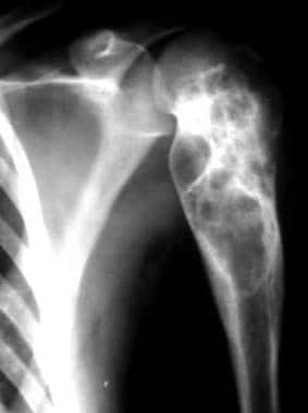 Aneurysmal bone cyst of the upper arm. Courtesy of Johannes Stahl, The Virtual Radiological Case Collection.
Jaffe and Lichtenstein first described ABC as its own entity in 1942, when they noted "a peculiar blood-containing cyst of large size." [9] Two cases were reported in which a lesion with a "soap-bubble" appearance on radiographs was found on the superior pubic ramus of a 17-year-old male and on the second vertebra of an 18-year-old male. The lesions were expansile and showed evidence of erosion of the surrounding bone and encroachment of the surrounding tissues. Upon surgical exposure of the lesions, a thin, bony wall that contained bloody fluid was found.
Jaffe and Lichtenstein suggested that ABCs may have been mistaken for other benign and malignant bone tumors in the past. [9] Although ABC is a separate entity, in some situations, distinguishing an ABC from a giant cell tumor of bone or a telangiectatic osteosarcoma is difficult.
The true etiology and pathophysiology remain a mystery, but the mainstay of treatment has been intralesional curettage. [10] Recurrence is not uncommon. [11, 12] Other surgical options include en-bloc resection or wide excision, selective arterial embolization, and curettage with locally applied adjuvants such as liquid nitrogen, argon beam photocoagulation, or phenol.
Anatomy
Because ABCs may affect any bone in the body, the relevant surgical anatomy necessarily varies with location. The long tubular bones are the most common sites for ABCs, followed by the spine and the flat bones. These three areas account for 80% of all ABCs. When present in long tubular bones, ABCs tend to be eccentrically located in the metaphysis.
ABCs least commonly involve a subperiosteal location, where they may form a predominant soft-tissue mass. However, ABCs can occur in any location, including the diaphysis and epiphysis.
Rarely, ABCs have also been known to affect an adjacent bone; however, spinal ABCs are associated with a higher incidence of contiguous lesions. Almost all ABCs of the spine involve the posterior elements, and a high incidence of neurologic symptoms is observed, as well as more local aggressive behavior.
The pelvis accounts for approximately 50% of lesions occurring in the flat bones. [13] Secondary lesions tend to have a predilection for the areas of the body in which the primary lesion typically arises.
In a published review of 897 cases of ABC, the following rates of occurrence were reported [14] :
-
Tibia – 17.5%
-
Femur – 15.9%
-
Vertebra – 11.2%
-
Pelvis – 11.6%
-
Humerus – 9.1%
-
Fibula – 7.3%
-
Foot – 6.3%
-
Hand – 4.7%
-
Ulna – 3.8%
-
Radius – 3.1%
-
Other – 9.2%
Pathophysiology
The true pathophysiology of ABCs is unknown. [10]
Different theories about several vascular malformations exist; these include arteriovenous fistulas (AVFs) and venous blockage. The vascular lesions then cause increased pressure, expansion, erosion, and resorption of the surrounding bone. The malformation is also believed to cause local hemorrhage that initiates the formation of reactive osteolytic tissue. Findings from a study in which manometric pressures within the ABCs were measured supported the theory of altered hemodynamics.
Most primary ABCs demonstrate a t(16;17)(q22;p13) fusion of the TRE17/CDH11-USP6 oncogene. This fusion leads to increased cellular cadherin-11 activity that seems to arrest osteoblastic maturation in a more primitive state. [15] This process may be the neoplastic driving force behind primary ABCs as opposed to secondary ABCs, which seem to occur reactively as a result of another underlying disease process. [16]
Etiology
The true etiology of ABCs is also unknown. Most investigators believe that ABCs are the result of a vascular malformation within the bone; however, the ultimate cause of the malformation is a topic of controversy. Three commonly proposed theories are as follows:
-
ABCs may be caused by a reaction secondary to another bony lesion - This theory has been proposed because of the high incidence of accompanying tumors in 23-32% of ABCs; although giant cell tumors of bone are most commonly present, many other benign and malignant tumors are found, including fibrous dysplasia, osteoblastoma, chondromyxoid fibroma, nonossifying fibroma, chondroblastoma, osteosarcoma, chondrosarcoma, unicameral or solitary bone cyst, hemangioendothelioma, and metastatic carcinoma; ABCs in the presence of another lesion are called secondary ABCs, and treatment of these ABCs is based on what is appropriate for the underlying tumor
-
ABCs may arise de novo; those that arise without evidence of another lesion are classified as primary ABCs
-
ABCs may arise in an area of previous trauma
A certain percentage of primary ABCs may be truly neoplastic—as opposed to vascular, developmental, or reactive—phenomena. It has been shown that as many as 69% of primary ABCs demonstrate a characteristic clonal t(16;17) genetic translocation [17] leading to upregulation of the TRE17/USP6 oncogene, [15, 16] whereas no secondary ABCs demonstrate this cytogenetic aberration.
Epidemiology
ABCs are generally considered rare, accounting for only 1-6% of all primary bony tumors. A group from Austria reported an annual incidence of 0.14 ABCs per 100,000 people [18] ; however, the true incidence is difficult to calculate because of the existence of spontaneous regression and clinically silent cases.
A biopsy-proven incidence study from the Netherlands showed that ABCs were the second most common tumor or tumorlike lesion found in children. [19]
Most studies have also found a slightly increased incidence in women. Although the ABC can appear in persons of any age, it is generally a disease of the young (albeit a rare one in the very young). About 50-70% of ABCs occur in the second decade of life, with 70-86% occurring in patients younger than 20 years. The mean patient age at onset is 13-17.7 years.
Prognosis
The prognosis for an ABC is generally excellent, though some patients need repeated treatments because of recurrence, which is the most common problem encountered when treating an ABC.
The overall cure rate is 90-95%. [20, 21] A younger age, open growth plates, and a metaphyseal location all have been associated with an increased risk of recurrence. [20] The stage of the ABC has not been shown to influence the rate of recurrence; however, most clinicians believe that Enneking/Musculoskeletal Tumor Society (MSTS) stage 3 lesions have the highest recurrence rate, other factors being equal. Capanna morphologic types I and II recur more often than types III, IV, and V.
Reported primary recurrence rates have varied greatly. Small studies have shown a benefit to using selective arterial embolization, and some authors have advocated it as a first-line treatment. Other authors have argued that not enough data on selective embolization exist and that surgery is the first-line treatment. Intralesional excision has the most data to suggest that it is a safe and effective method.
Recurrence rates for different techniques have varied. Some studies have reported recurrence rates as high as 59% with intralesional excision [22] and as low as 0% with resection. [14] In a summary of studies of different treatment methods, the following rates of recurrence were reported [14] :
-
Irradiation – 34 performed with 4 recurrences (11.8% recurrence rate)
-
Irradiation and curettage – 35 performed with 5 recurrences (14.3% recurrence rate)
-
Curettage and bone graft – 484 performed with 149 recurrences (30.8% recurrence rate)
-
Curettage and cryobiopsy – 78 performed with 10 recurrences (12.8% recurrence rate)
-
Marginal resection – 81 performed with 6 recurrences (7.4% recurrence rate)
-
Wide resection – 59 performed with 0 recurrences (0% recurrence rate)
A systematic review and meta-analysis by Cruz et al assessed recurrence rates after percutaneous treatment of primary ABCs, comparing selective arterial embolization with sclerotherapy. [23] For percutaneous treatment overall, the average success rate was 91.11% (37 lesion recurrences in 416 patients). However, the recurrence rate in patients treated with sclerotherapy was 9%, compared with 19% in those treated with selective arterial embolization.
-
Aneurysmal bone cyst of the upper arm. Courtesy of Johannes Stahl, The Virtual Radiological Case Collection.
-
Aneurysmal bone cyst of the upper arm. Courtesy of Johannes Stahl, The Virtual Radiological Case Collection.
-
Aneurysmal bone cyst affecting the lumbar spine. Courtesy of Nick Oldnall, Index: Spine.
-
Aneurysmal bone cyst of the upper arm in a 13-year-old female adolescent. Left to right, the radiographs were obtained over a period of approximately 2 months. Courtesy of Armed Forces Institute of Pathology.
-
Aneurysmal bone cyst of the upper arm in an 18-year-old woman. Note the cortical erosion and slight expansion. Courtesy of Armed Forces Institute of Pathology.
-
Aneurysmal bone cyst of the upper arm. Used with permission from the American Registry of Pathology. Courtesy of Armed Forces Institute of Pathology.
-
Aneurysmal bone cyst of the upper arm. Used with permission from the American Registry of Pathology. Courtesy of Armed Forces Institute of Pathology.
-
In this histologic image, the vascular spaces vary widely in size and shape, and the septa range from thin to broad. Well-formed bone is focally present. Courtesy of Armed Forces Institute of Pathology.
-
In this histologic image, the septum contains fibroblasts, mononuclear histiocytelike cells, multinucleated giant cells, and capillaries. The lining of the large vascular spaces may be indistinct or consist of a single layer of attenuated stromal cells, as seen here. Used with permission from the American Registry of Pathology. Courtesy of Armed Forces Institute of Pathology.
-
This histologic image demonstrates lesional tissue that abuts the striated tissue where the cortex has been completely destroyed. Used with permission from the American Registry of Pathology. Courtesy of Armed Forces Institute of Pathology.
-
This histologic image shows a fibrous septum that contains a long strand of osteoid that appears to have buckled in the center. Used with permission from the American Registry of Pathology. Courtesy of Armed Forces Institute of Pathology.
-
Aneurysmal bone cyst of distal radius in 11-year-old male. Note multiple septations, lytic cyst cavity, and extensive cortical thinning and expansion wider than adjacent distal radius physis in both anteroposterior and lateral planes.
-
Axial and sagittal T2-weighted MRI of distal femoral aneurysmal bone cyst. Note multiple fluid-fluid levels throughout lesion consistent with multiple blood-filled cavities separated by small septa.


