Semi-Solid Pharmaceutical Formulations for the Delivery of Papain Nanoparticles


Abstract
:1. Introduction
2. Materials and Methods
2.1. Materials
2.2. Methods
2.2.1. Nanoparticles Synthesis
2.2.2. Development of Semi-Solid Formulations
2.2.3. Papain Loading
2.2.4. Characterization
Nanoparticles Size
Nanoparticles Crosslinking
Stability Assessment
Scanning Electron Microscopy
Protein Distribution
Proteolytic Activity in the Gel Formulation
Papain Release
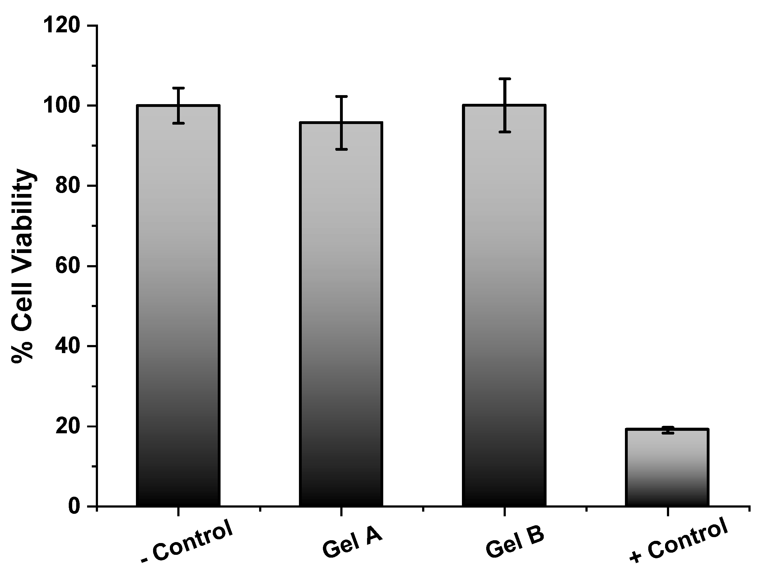
Cytotoxicity
3. Results and Discussion
3.1. Nanoparticles
3.2. Semi-Solid Formulation Stability Assessment
4. Conclusions
Author Contributions
Funding
Acknowledgments
Conflicts of Interest
References
- Homaei, A.; Sariri, R.; Vianello, F.; Stevanato, R. Enzyme immobilization: An update. J. Chem. Biol. 2013, 6, 185–205. [Google Scholar] [CrossRef] [PubMed] [Green Version]
- Kamphuis, I.; Kalk, K.; Swarte, M.; Drenth, J. Structure of papain refined at 1.65 Å resolution. J. Mol. Biol. 1984, 179, 233–256. [Google Scholar] [CrossRef]
- Dos Anjos, M.M.; Da Silva, A.A.; De Pascoli, I.C.; Mikcha, J.M.G.; Machinski, M.; Peralta, R.M.; Filho, B.A.D.A. Antibacterial activity of papain and bromelain on Alicyclobacillus spp. Int. J. Food Microbiol. 2016, 216, 121–126. [Google Scholar] [CrossRef] [PubMed]
- Da Silva, C.R.; Oliveira, M.B.N.; Motta, E.S.; De Almeida, G.S.; Varanda, L.L.; De Pádula, M.; Leitão, Á.C.; Caldeira-De-Araujo, A. Genotoxic and Cytotoxic Safety Evaluation of Papain (Carica papaya L.) Using In Vitro Assays. J. Biomed. Biotechnol. 2010, 2010, 197898. [Google Scholar] [CrossRef] [PubMed] [Green Version]
- Beuth, J. Proteolytic Enzyme Therapy in Evidence-Based Complementary Oncology: Fact or Fiction? Integr. Cancer Ther. 2008, 7, 311–316. [Google Scholar] [CrossRef]
- Müller, A.; Barat, S.; Chen, X.; Bui, K.C.; Bozko, P.; Malek, N.P.; Plentz, R.R. Comparative study of antitumor effects of bromelain and papain in human cholangiocarcinoma cell lines. Int. J. Oncol. 2016, 48, 2025–2034. [Google Scholar] [CrossRef] [Green Version]
- Sim, Y.-C.; Lee, S.-G.; Lee, D.-C.; Kang, B.-Y.; Park, K.-M.; Lee, J.-Y.; Kim, M.-S.; Chang, I.-S.; Rhee, J.-S. Stabilization of papain and lysozyme for application to cosmetic products. Biotechnol. Lett. 2000, 22, 137–140. [Google Scholar] [CrossRef]
- Arnold, F.H. Protein engineering for unusual environments. Curr. Opin. Biotechnol. 1993, 4, 450–455. [Google Scholar] [CrossRef]
- Atacan, K.; Özacar, M.; Özacar, M. Investigation of antibacterial properties of novel papain immobilized on tannic acid modified Ag/CuFe2O4 magnetic nanoparticles. Int. J. Biol. Macromol. 2018, 109, 720–731. [Google Scholar] [CrossRef]
- FDA Topical Drug Products Containing Papain. Available online: https://www.govinfo.gov/content/pkg/FR-2008-09-23/pdf/E8-22300.pdf (accessed on 27 November 2020).
- Chapman, R.; Stenzel, M.H. All Wrapped up: Stabilization of Enzymes within Single Enzyme Nanoparticles. J. Am. Chem. Soc. 2019, 141, 2754–2769. [Google Scholar] [CrossRef]
- Varca, G.H.C.; Ferraz, C.C.; Lopes, P.S.; Mathor, M.B.; Grasselli, M.; Lugao, A.B. Radio-synthesized protein-based nanoparticles for biomedical purposes. Radiat. Phys. Chem. 2014, 94, 181–185. [Google Scholar] [CrossRef]
- Varca, G.H.; Kadlubowski, S.; Wolszczak, M.; Lugão, A.; Rosiak, J.; Ulanski, P. Synthesis of papain nanoparticles by electron beam irradiation ⿿ A pathway for controlled enzyme crosslinking. Int. J. Biol. Macromol. 2016, 92, 654–659. [Google Scholar] [CrossRef] [PubMed]
- Fazolin, G.N.; Varca, G.H.C.; Kadlubowski, S.; Sowinski, S.; Lugao, A.B. The effects of radiation and experimental conditions over papain nanoparticle formation: Towards a new generation synthesis. Radiat. Phys. Chem. 2020, 169, 107984. [Google Scholar] [CrossRef]
- Lemos, C.N.; Pereira, F.; Dalmolin, L.F.; Cubayachi, C.; Ramos, D.N.; Lopez, R.F.V. Nanoparticles Influence in Skin Penetration of Drugs: In Vitro and In Vivo Characterization. In Nanostructures for the Engineering of Cells, Tissues and Organs; William Andrew Publishing: Oxford, UK, 2018; ISBN 9780128136669. [Google Scholar]
- Budama-Kilinc, Y.; Cakir-Koc, R.; Kecel-Gunduz, S.; Zorlu, T.; Kokcu, Y.; Bicak, B.; Karavelioglu, Z.; Ozel, A.E. Papain Loaded Poly(ε-Caprolactone) Nanoparticles: In-silico and In-Vitro Studies. J. Fluoresc. 2018, 28, 1127–1142. [Google Scholar] [CrossRef]
- Chen, Y.-Y.; Lu, Y.-H.; Ma, C.-H.; Tao, W.-W.; Zhu, J.-J.; Zhang, X. A novel elastic liposome for skin delivery of papain and its application on hypertrophic scar. Biomed. Pharmacother. 2017, 87, 82–91. [Google Scholar] [CrossRef]
- Feksa, L.R.; Troian, E.A.; Muller, C.D.; Viegas, F.; Machado, A.B.; Rech, V.C. Hydrogels for biomedical applications. In Nanostructures for the Engineering of Cells, Tissues and Organs; William Andrew Publishing: Norwich, NY, USA, 2018; Volume 54, pp. 403–438. [Google Scholar] [CrossRef]
- Namazi, H.; Rakhshaei, R.; Hamishehkar, H.; Kafil, H.S. Antibiotic loaded carboxymethylcellulose/MCM-41 nanocomposite hydrogel films as potential wound dressing. Int. J. Biol. Macromol. 2016, 85, 327–334. [Google Scholar] [CrossRef]
- Yadollahi, M.; Gholamali, I.; Namazi, H.; Aghazadeh, M. Synthesis and characterization of antibacterial carboxymethylcellulose/CuO bio-nanocomposite hydrogels. Int. J. Biol. Macromol. 2015, 73, 109–114. [Google Scholar] [CrossRef]
- Gaaz, T.S.; Sulong, A.B.; Akhtar, M.N.; Kadhum, A.A.H.; Mohamad, A.B.; Al-Amiery, A.A. Properties and Applications of Polyvinyl Alcohol, Halloysite Nanotubes and Their Nanocomposites. Molecules 2015, 20, 22833–22847. [Google Scholar] [CrossRef] [Green Version]
- Peppas, N.A.; Mongia, N.K. Ultrapure poly(vinyl alcohol) hydrogels with mucoadhesive drug delivery characteristics. Eur. J. Pharm. Biopharm. 1997, 43, 51–58. [Google Scholar] [CrossRef]
- Rac, V.; Lević, S.; Balanč, B.D.; Graells, B.O.; Bijelić, G. PVA Cryogel as model hydrogel for iontophoretic transdermal drug delivery investigations. Comparison with PAA/PVA and PAA/PVP interpenetrating networks. Colloids Surf. B Biointerfaces 2019, 180, 441–448. [Google Scholar] [CrossRef]
- Sebe, I. (PhD hallgató); Ostorhazi, E.; Fekete, A.; Kovacs, K.N.; Zelko, R.; Kovalszky, I.; Li, W.; Wade, J.D.; Szabo, D.; Otvos, L. Polyvinyl alcohol nanofiber formulation of the designer antimicrobial peptide APO sterilizes Acinetobacter baumannii-infected skin wounds in mice. Amino Acids 2015, 48, 203–211. [Google Scholar] [CrossRef]
- Timofejeva, A.; D’Este, M.; Loca, D. Calcium phosphate/polyvinyl alcohol composite hydrogels: A review on the freeze-thawing synthesis approach and applications in regenerative medicine. Eur. Polym. J. 2017, 95, 547–565. [Google Scholar] [CrossRef]
- Ghorpade, V.S.; Dias, R.J.; Mali, K.K.; Mulla, S.I. Citric acid crosslinked carboxymethylcellulose-polyvinyl alcohol hydrogel films for extended release of water soluble basic drugs. J. Drug Deliv. Sci. Technol. 2019, 52, 421–430. [Google Scholar] [CrossRef]
- Wang, S.; Zhang, Q.; Tan, B.; Liu, L.; Shi, L. pH-Sensitive Poly(Vinyl Alcohol)/Sodium Carboxymethylcellulose Hydrogel Beads for Drug Delivery. J. Macromol. Sci. Part B 2011, 50, 2307–2317. [Google Scholar] [CrossRef]
- Fazolin, G.N.; Varca, G.H.; De Freitas, L.F.; Rokita, B.; Kadlubowski, S.; Lugão, A.B. Simultaneous intramolecular crosslinking and sterilization of papain nanoparticles by gamma radiation. Radiat. Phys. Chem. 2020, 171, 108697. [Google Scholar] [CrossRef]
- ISO 22412:2017. Particle Size Analysis—Dynamic Ligh Scattering (DLS); ISO: Geneva, Switzerland, 2017. [Google Scholar]
- ANVISA. Guia de Estabilidade dos Cosméticos Anvisa; ANVISA: Brasília, Brazil, 2004; Volume 1, ISBN 85-88233-15-0.
- Bradford, M.M. A Rapid and Sensitive Method for the Quantitation Microgram Quantities of Protein Utilizing the Principle of Protein-Dye Binding. Anal. Biochem. 1976, 72, 248–254. [Google Scholar] [CrossRef]
- Ferraz, C.C.; Varca, G.H.C.; Vila, M.M.D.C.; Lopes, P.S. Validation of in vitro analytical method to measure papain activity in pharmaceutical formulations. Int. J. Pharm. Pharm. Sci. 2014, 6, 658–661. [Google Scholar]
- Varca, G.H.; Perossi, G.G.; Grasselli, M.; Lugao, A.B. Radiation synthesized protein-based nanoparticles: A technique overview. Radiat. Phys. Chem. 2014, 105, 48–52. [Google Scholar] [CrossRef]
- Jon; Amri, E.; Mamboya, F. Papain, a Plant Enzyme of Biological Importance: A Review. Am. J. Biochem. Biotechnol. 2012, 8, 99–104. [Google Scholar] [CrossRef] [Green Version]
- Uhlig, H. Industrial Enzymes and their Applications, 1st ed.; Wiley-Interscience: New York, NY, USA, 1998. [Google Scholar]
- Ferraz, C.C.; Varca, G.H.; Lopes, P.S.; Mathor, M.B.; Lugao, A.B. Radio-synthesized polyacrylamide hydrogels for proteins release. Radiat. Phys. Chem. 2014, 94, 186–189. [Google Scholar] [CrossRef]


| Sample | Size (d.nm) | Polydispersity Index |
Enzymatic Activity |
|---|---|---|---|
| Nanopapain | 7.0 ± 0.1 | 0.34 | 88.5% ± 7.39 |
| Native papain | 3.3 ± 0.4 | 0.39 | 100% ± 1.03 |
| Formulation | Content | Centrifugation | Aspect | Color | Smell |
|---|---|---|---|---|---|
| Gel A | 0.2% PVA 2% CMC |
stable | Homogeneous and translucent | Colorless | No change |
| Gel A + papain (control) | Gel A + 0.2% native papain | stable | Homogeneous and translucent | Colorless | No change |
| Gel A + nanopapain | Gel A + 0.2% nanopapain | stable | Homogeneous and translucent | Colorless | No change |
| Gel B | 0.2% PVA 3% CMC |
stable | Homogeneous and translucent | Colorless | No change |
| Gel B + papain (control) | Gel B + 0.2% native papain | stable | Homogeneous and translucent | Colorless | No change |
| Gel B + nanopapain | Gel B + 0.2% nanopapain | stable | Homogeneous and translucent | Colorless | No change |
|
Publisher’s Note: MDPI stays neutral with regard to jurisdictional claims in published maps and institutional affiliations.
|
© 2020 by the authors. Licensee MDPI, Basel, Switzerland. This article is an open access article distributed under the terms and conditions of the Creative Commons Attribution (CC BY) license (http://creativecommons.org/licenses/by/4.0/).
Share and Cite
Lima, C.S.A.d.; Varca, J.P.R.O.; Nogueira, K.M.; Fazolin, G.N.; Freitas, L.F.d.; Souza, E.W.d.; Lugão, A.B.; Varca, G.H.C. Semi-Solid Pharmaceutical Formulations for the Delivery of Papain Nanoparticles. Pharmaceutics 2020, 12, 1170. https://doi.org/10.3390/pharmaceutics12121170
Lima CSAd, Varca JPRO, Nogueira KM, Fazolin GN, Freitas LFd, Souza EWd, Lugão AB, Varca GHC. Semi-Solid Pharmaceutical Formulations for the Delivery of Papain Nanoparticles. Pharmaceutics. 2020; 12(12):1170. https://doi.org/10.3390/pharmaceutics12121170
Chicago/Turabian StyleLima, Caroline S. A. de, Justine P. R. O. Varca, Kamila M. Nogueira, Gabriela N. Fazolin, Lucas F. de Freitas, Eliseu W. de Souza, Ademar B. Lugão, and Gustavo. H. C. Varca. 2020. "Semi-Solid Pharmaceutical Formulations for the Delivery of Papain Nanoparticles" Pharmaceutics 12, no. 12: 1170. https://doi.org/10.3390/pharmaceutics12121170