


Viruses 2024, 16(5), 678; https://doi.org/10.3390/v16050678 (registering DOI) - 25 Apr 2024
Abstract
Porcine reproductive and respiratory syndrome virus (PRRSV), a member of the Arteriviridae family, represents a persistent menace to the global pig industry, causing reproductive failure and respiratory disease in pigs. In this study, we delved into the role of histone deacetylases (HDAC2) during [...] Read more.
Porcine reproductive and respiratory syndrome virus (PRRSV), a member of the Arteriviridae family, represents a persistent menace to the global pig industry, causing reproductive failure and respiratory disease in pigs. In this study, we delved into the role of histone deacetylases (HDAC2) during PRRSV infection. Our findings revealed that HDAC2 expression is downregulated upon PRRSV infection. Notably, suppressing HDAC2 activity through specific small interfering RNA led to an increase in virus production, whereas overexpressing HDAC2 effectively inhibited PRRSV replication by boosting the expression of IFN-regulated antiviral molecules. Furthermore, we identified the virus’s nonstructural protein 11 (nsp11) as a key player in reducing HDAC2 levels. Mutagenic analyses of PRRSV nsp11 revealed that its antagonistic effect on the antiviral activity of HDAC2 is dependent on its endonuclease activity. In summary, our research uncovered a novel immune evasion mechanism employed by PRRSV, providing crucial insights into the pathogenesis of this virus and guiding the development of innovative prevention strategies against PRRSV infection. Full article
(This article belongs to the Section Viral Immunology, Vaccines, and Antivirals)
► Show Figures
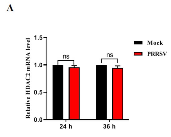
Figure 1


